 Movie
Movie Controller

[English] 日本語
 Yorodumi
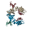
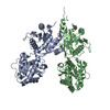
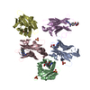
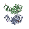
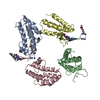
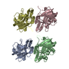
- PDB-1fcc: CRYSTAL STRUCTURE OF THE C2 FRAGMENT OF STREPTOCOCCAL PROTEIN G I... -
+ Open data

- Basic information
| Entry | Database: PDB / ID: 1fcc | ||||||
|---|---|---|---|---|---|---|---|
| Title | CRYSTAL STRUCTURE OF THE C2 FRAGMENT OF STREPTOCOCCAL PROTEIN G IN COMPLEX WITH THE FC DOMAIN OF HUMAN IGG | ||||||
 Components Components |
| ||||||
 Keywords Keywords | COMPLEX (ANTIBODY/ANTIGEN) / COMPLEX (ANTIBODY-ANTIGEN) / COMPLEX (ANTIBODY-ANTIGEN) complex | ||||||
| Function / homology |  Function and homology information Function and homology informationcomplement-dependent cytotoxicity / antibody-dependent cellular cytotoxicity / Fc-gamma receptor I complex binding / IgG binding / immunoglobulin complex, circulating / Classical antibody-mediated complement activation / immunoglobulin receptor binding / Initial triggering of complement / IgG immunoglobulin complex / FCGR activation ...complement-dependent cytotoxicity / antibody-dependent cellular cytotoxicity / Fc-gamma receptor I complex binding / IgG binding / immunoglobulin complex, circulating / Classical antibody-mediated complement activation / immunoglobulin receptor binding / Initial triggering of complement / IgG immunoglobulin complex / FCGR activation / complement activation, classical pathway / Role of phospholipids in phagocytosis / antigen binding / FCGR3A-mediated IL10 synthesis / Regulation of Complement cascade / B cell receptor signaling pathway / FCGR3A-mediated phagocytosis / Regulation of actin dynamics for phagocytic cup formation / antibacterial humoral response / Interleukin-4 and Interleukin-13 signaling / blood microparticle / adaptive immune response / : / extracellular exosome / extracellular region / plasma membrane Similarity search - Function | ||||||
| Biological species |  Homo sapiens (human) Homo sapiens (human) Streptococcus (bacteria) Streptococcus (bacteria) | ||||||
| Method |  X-RAY DIFFRACTION / Resolution: 3.2 Å X-RAY DIFFRACTION / Resolution: 3.2 Å | ||||||
 Authors Authors | Sauer-Eriksson, A.E. / Kleywegt, G.J. / Uhlen, M. / Jones, T.A. | ||||||
 Citation Citation | Journal: Structure / Year: 1995 Title: Crystal structure of the C2 fragment of streptococcal protein G in complex with the Fc domain of human IgG. Authors: Sauer-Eriksson, A.E. / Kleywegt, G.J. / Uhlen, M. / Jones, T.A. | ||||||
| History |
|
- Structure visualization
| Structure viewer | Molecule: MolmilJmol/JSmol |
|---|
- Downloads & links
-Download
| PDBx/mmCIF format | 1fcc.cif.gz | 102.1 KB | Display | PDBx/mmCIF format |
|---|---|---|---|---|
| PDB format | pdb1fcc.ent.gz | 80 KB | Display | PDB format |
| PDBx/mmJSON format | 1fcc.json.gz | Tree view | PDBx/mmJSON format | |
| Others |  Other downloads Other downloads |
-Validation report
| Arichive directory | https://data.pdbj.org/pub/pdb/validation_reports/fc/1fccftp://data.pdbj.org/pub/pdb/validation_reports/fc/1fcc | HTTPS FTP |
|---|
-Related structure data
| Similar structure data |
|---|


-Links
 PDBj
PDBj


- Assembly
| Deposited unit | 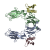
| ||||||||
|---|---|---|---|---|---|---|---|---|---|
| 1 |
| ||||||||
| Unit cell |
| ||||||||
| Atom site foot note | 1: CIS PROLINE - PRO A 374 | ||||||||
| Noncrystallographic symmetry (NCS) | NCS oper: (Code: given Matrix: (-0.700155, -0.700538, -0.138305), Vector: Details | MTRIX THE TRANSFORMATIONS PRESENTED ON MTRIX RECORDS BELOW DESCRIBE NON-CRYSTALLOGRAPHIC RELATIONSHIPS AMONG THE VARIOUS DOMAINS IN THIS ENTRY. APPLYING THE APPROPRIATE MTRIX TRANSFORMATION TO THE RESIDUES LISTED IN THIS ENTRY WILL YIELD APPROXIMATE COORDINATES FOR THE RESIDUES OF THE OTHER MONOMERS IN THE ASYMMETRIC UNIT NOT PRESENTED IN THIS ENTRY. | |
-Components
| #1: Protein | Mass: 23519.654 Da / Num. of mol.: 2 Source method: isolated from a genetically manipulated source Source: (gene. exp.) Homo sapiens (human) / Cell line: HYBRIDOMA / Gene: N-TERMINAL FRAGMENT OF / Plasmid: PEB2ZHIS GENE: N-TERMINAL FRAGMENT OF / Production host: #2: Protein | Mass: 6157.665 Da / Num. of mol.: 2 Source method: isolated from a genetically manipulated source Source: (gene. exp.) Streptococcus (bacteria) / Genus: Streptococcus / Cell line: HYBRIDOMA / Gene: N-TERMINAL FRAGMENT OF / Plasmid: PEB2ZHIS GENE: N-TERMINAL FRAGMENT OF / Production host: Compound details | FIVE AMINO ACIDS DIFFER IN THEIR AMIDATION STATES AND TWO HAVE THE ALLOTYPIC MARKER: E119, M121. | Has protein modification | Y | |
|---|
-Experimental details
-Experiment
| Experiment | Method: X-RAY DIFFRACTION |
|---|
- Sample preparation
| Crystal | Density Matthews: 4.13 Å3/Da / Density % sol: 70.2 % |
|---|---|
| Crystal grow | *PLUS Method: vapor diffusion, hanging dropDetails: Jancarik, J., (1991) J. Appl. Crystallogr., 24, 409. |
| Components of the solutions | *PLUS Conc.: 10 mg/ml / Common name: protein |
-Data collection
| Radiation | Scattering type: x-ray |
|---|---|
| Radiation wavelength | Relative weight: 1 |
| Reflection | Num. obs: 12297 / % possible obs: 72 % |
| Reflection | *PLUS Highest resolution: 3.2 Å / Lowest resolution: 9999 Å / Num. measured all: 41581 / Rmerge(I) obs: 0.089 |
- Processing
| Software |
| ||||||||||||||||||||||||||||||||||||||||||||||||||||||||||||
|---|---|---|---|---|---|---|---|---|---|---|---|---|---|---|---|---|---|---|---|---|---|---|---|---|---|---|---|---|---|---|---|---|---|---|---|---|---|---|---|---|---|---|---|---|---|---|---|---|---|---|---|---|---|---|---|---|---|---|---|---|---|
| Refinement | Resolution: 3.2→8 Å
| ||||||||||||||||||||||||||||||||||||||||||||||||||||||||||||
| Displacement parameters | Biso mean: 41.6 Å2 | ||||||||||||||||||||||||||||||||||||||||||||||||||||||||||||
| Refinement step | Cycle: LAST / Resolution: 3.2→8 Å
| ||||||||||||||||||||||||||||||||||||||||||||||||||||||||||||
| Refine LS restraints |
| ||||||||||||||||||||||||||||||||||||||||||||||||||||||||||||
| Refinement | *PLUS Highest resolution: 3.5 Å | ||||||||||||||||||||||||||||||||||||||||||||||||||||||||||||
| Solvent computation | *PLUS | ||||||||||||||||||||||||||||||||||||||||||||||||||||||||||||
| Displacement parameters | *PLUS | ||||||||||||||||||||||||||||||||||||||||||||||||||||||||||||
| Refine LS restraints | *PLUS
|
