 Movie
Movie Controller


+ Open data

- Basic information
| Entry | Database: PDB / ID: 2pqs | ||||||
|---|---|---|---|---|---|---|---|
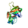
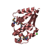
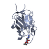
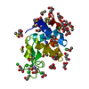
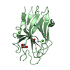
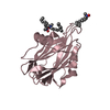
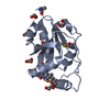
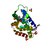
| Title | Crystal Structure of the Bovine Lactadherin C2 Domain | ||||||
 Components Components | Lactadherin | ||||||
 Keywords Keywords | CELL ADHESION / C2 domain / lactadherin / membrane binding | ||||||
| Function / homology |  Function and homology information Function and homology informationacrosomal membrane / phosphatidylserine binding / single fertilization / angiogenesis / cell adhesion / : Similarity search - Function | ||||||
| Biological species |  | ||||||
| Method |  X-RAY DIFFRACTION / SYNCHROTRON / MOLECULAR REPLACEMENT / Resolution: 2.4 Å X-RAY DIFFRACTION / SYNCHROTRON / MOLECULAR REPLACEMENT / Resolution: 2.4 Å | ||||||
 Authors Authors | Huang, M. / Furie, B.C. | ||||||
 Citation Citation | Journal: J.Mol.Biol. / Year: 2007 Title: Crystal structure of the bovine lactadherin C2 domain, a membrane binding motif, shows similarity to the C2 domains of factor V and factor VIII. Authors: Lin, L. / Huai, Q. / Huang, M. / Furie, B. / Furie, B.C. | ||||||
| History |
|
- Structure visualization
| Structure viewer | Molecule: MolmilJmol/JSmol |
|---|
- Downloads & links
-Download
| PDBx/mmCIF format | 2pqs.cif.gz | 136 KB | Display | PDBx/mmCIF format |
|---|---|---|---|---|
| PDB format | pdb2pqs.ent.gz | 108.6 KB | Display | PDB format |
| PDBx/mmJSON format | 2pqs.json.gz | Tree view | PDBx/mmJSON format | |
| Others |  Other downloads Other downloads |
-Validation report
| Arichive directory | https://data.pdbj.org/pub/pdb/validation_reports/pq/2pqsftp://data.pdbj.org/pub/pdb/validation_reports/pq/2pqs | HTTPS FTP |
|---|
-Related structure data
| Similar structure data |
|---|


-Links
 PDBj
PDBj


- Assembly
| Deposited unit | 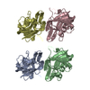
| ||||||||
|---|---|---|---|---|---|---|---|---|---|
| 1 | 
| ||||||||
| 2 | 
| ||||||||
| 3 | 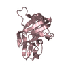
| ||||||||
| 4 | 
| ||||||||
| Unit cell |
|
-Components
| #1: Protein | Mass: 18095.232 Da / Num. of mol.: 4 / Fragment: C2 domain (Residues 270-427) Source method: isolated from a genetically manipulated source Source: (gene. exp.)  Pichia pastoris (fungus) / References: UniProt: Q95114 Pichia pastoris (fungus) / References: UniProt: Q95114#2: Water | ChemComp-HOH / |  Mass: 18.015 Da / Num. of mol.: 119 / Source method: isolated from a natural source / Formula: H2O Mass: 18.015 Da / Num. of mol.: 119 / Source method: isolated from a natural source / Formula: H2OHas protein modification | Y | |
|---|
-Experimental details
-Experiment
| Experiment | Method: X-RAY DIFFRACTION / Number of used crystals: 1 |
|---|
- Sample preparation
| Crystal | Density Matthews: 3.33 Å3/Da / Density % sol: 63.06 % |
|---|---|
| Crystal grow | Temperature: 295 K / Method: evaporation / pH: 7.5 Details: 15% PEG4K, 20% glycerol, 50 mM HEPES, pH 7.5, EVAPORATION, temperature 295K |
-Data collection
| Diffraction | Mean temperature: 100 K |
|---|---|
| Diffraction source | Source: SYNCHROTRON / Site: NSLS  / Beamline: X12C / Wavelength: 1 Å / Beamline: X12C / Wavelength: 1 Å |
| Detector | Detector: IMAGE PLATE |
| Radiation | Monochromator: Si(111) channel-cut crystal monochromator / Protocol: SINGLE WAVELENGTH / Monochromatic (M) / Laue (L): M / Scattering type: x-ray |
| Radiation wavelength | Wavelength: 1 Å / Relative weight: 1 |
| Reflection | Resolution: 2.4→50 Å / Num. all: 19426 / Num. obs: 19057 / % possible obs: 98.1 % / Observed criterion σ(F): 0 / Observed criterion σ(I): 0 / Redundancy: 8 % / Biso Wilson estimate: 39.9 Å2 / Rmerge(I) obs: 0.06 / Net I/σ(I): 30.2 |
| Reflection shell | Resolution: 2.4→2.49 Å / Redundancy: 7.8 % / Rmerge(I) obs: 0.353 / Mean I/σ(I) obs: 3.7 |
- Processing
| Software |
| ||||||||||||||||||||||||||||||||||||||||||||||||||||||||||||||||||||||||||||||||
|---|---|---|---|---|---|---|---|---|---|---|---|---|---|---|---|---|---|---|---|---|---|---|---|---|---|---|---|---|---|---|---|---|---|---|---|---|---|---|---|---|---|---|---|---|---|---|---|---|---|---|---|---|---|---|---|---|---|---|---|---|---|---|---|---|---|---|---|---|---|---|---|---|---|---|---|---|---|---|---|---|---|
| Refinement | Method to determine structure: MOLECULAR REPLACEMENT / Resolution: 2.4→45.26 Å / Rfactor Rfree error: 0.008 / Data cutoff high absF: 1171545.97 / Data cutoff low absF: 0 / Isotropic thermal model: RESTRAINED / Cross valid method: THROUGHOUT / σ(F): 0 / Stereochemistry target values: Engh & Huber
| ||||||||||||||||||||||||||||||||||||||||||||||||||||||||||||||||||||||||||||||||
| Solvent computation | Solvent model: FLAT MODEL / Bsol: 31.6315 Å2 / ksol: 0.339252 e/Å3 | ||||||||||||||||||||||||||||||||||||||||||||||||||||||||||||||||||||||||||||||||
| Displacement parameters | Biso mean: 44.4 Å2
| ||||||||||||||||||||||||||||||||||||||||||||||||||||||||||||||||||||||||||||||||
| Refine analyze |
| ||||||||||||||||||||||||||||||||||||||||||||||||||||||||||||||||||||||||||||||||
| Refinement step | Cycle: LAST / Resolution: 2.4→45.26 Å
| ||||||||||||||||||||||||||||||||||||||||||||||||||||||||||||||||||||||||||||||||
| Refine LS restraints |
| ||||||||||||||||||||||||||||||||||||||||||||||||||||||||||||||||||||||||||||||||
| LS refinement shell | Resolution: 2.4→2.55 Å / Rfactor Rfree error: 0.025 / Total num. of bins used: 6
| ||||||||||||||||||||||||||||||||||||||||||||||||||||||||||||||||||||||||||||||||
| Xplor file |
|
