 Movie
Movie Controller

[English] 日本語
 Yorodumi
- PDB-2onq: Gbeta1 stabilization by in vitro evolution and computational design -
+ Open data

- Basic information
| Entry | Database: PDB / ID: 2onq | ||||||
|---|---|---|---|---|---|---|---|
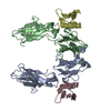
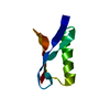
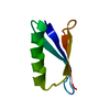
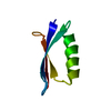
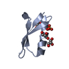
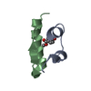
| Title | Gbeta1 stabilization by in vitro evolution and computational design | ||||||
 Components Components | Immunoglobulin G-binding protein G | ||||||
 Keywords Keywords | PROTEIN BINDING / beta sheet / alpha helix / improved hydrophobic packing of core residues | ||||||
| Function / homology |  Function and homology information Function and homology information | ||||||
| Biological species |  Streptococcus sp. (bacteria) Streptococcus sp. (bacteria) | ||||||
| Method |  X-RAY DIFFRACTION / MOLECULAR REPLACEMENT / Resolution: 1.7 Å X-RAY DIFFRACTION / MOLECULAR REPLACEMENT / Resolution: 1.7 Å | ||||||
 Authors Authors | Max, K.E.A. / Heinemann, U. | ||||||
 Citation Citation | Journal: J.Mol.Biol. / Year: 2007 Title: Optimization of the gbeta1 domain by computational design and by in vitro evolution: structural and energetic basis of stabilization. Authors: Wunderlich, M. / Max, K.E. / Roske, Y. / Mueller, U. / Heinemann, U. / Schmid, F.X. | ||||||
| History |
| ||||||
| Remark 300 | BIOMOLECULE: 1 THIS ENTRY CONTAINS THE CRYSTALLOGRAPHIC ASYMMETRIC UNIT WHICH CONSISTS OF 1 CHAIN(S) ...BIOMOLECULE: 1 THIS ENTRY CONTAINS THE CRYSTALLOGRAPHIC ASYMMETRIC UNIT WHICH CONSISTS OF 1 CHAIN(S). AUTHORS STATE THAT the biological unit information is unknown for this isolated domain. |
- Structure visualization
| Structure viewer | Molecule: MolmilJmol/JSmol |
|---|
- Downloads & links
-Download
| PDBx/mmCIF format | 2onq.cif.gz | 35.7 KB | Display | PDBx/mmCIF format |
|---|---|---|---|---|
| PDB format | pdb2onq.ent.gz | 24.7 KB | Display | PDB format |
| PDBx/mmJSON format | 2onq.json.gz | Tree view | PDBx/mmJSON format | |
| Others |  Other downloads Other downloads |
-Validation report
| Arichive directory | https://data.pdbj.org/pub/pdb/validation_reports/on/2onqftp://data.pdbj.org/pub/pdb/validation_reports/on/2onq | HTTPS FTP |
|---|
-Related structure data
| Related structure data | 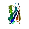 2on8C 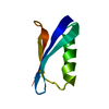 1pgaS C: citing same article ( S: Starting model for refinement |
|---|---|
| Similar structure data |


-Links
 PDBj
PDBj

- Assembly
| Deposited unit | 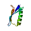
| ||||||||
|---|---|---|---|---|---|---|---|---|---|
| 1 |
| ||||||||
| Unit cell |
|
-Components
| #1: Antibody | Mass: 6326.997 Da / Num. of mol.: 1 / Fragment: residues 373-427 / Mutation: T2Q, Y3F, L7I, T16I, T18I, T25E, V29F, V39I Source method: isolated from a genetically manipulated source Source: (gene. exp.) Streptococcus sp. (bacteria) / Strain: G148 / Gene: spg / Plasmid: pET11a / Species (production host): Escherichia coli / Production host: |
|---|---|
| #2: Water | ChemComp-HOH /  Mass: 18.015 Da / Num. of mol.: 34 / Source method: isolated from a natural source / Formula: H2O Mass: 18.015 Da / Num. of mol.: 34 / Source method: isolated from a natural source / Formula: H2O |
-Experimental details
-Experiment
| Experiment | Method: X-RAY DIFFRACTION / Number of used crystals: 1 |
|---|
- Sample preparation
| Crystal | Density Matthews: 1.91 Å3/Da / Density % sol: 35.7 % |
|---|---|
| Crystal grow | Temperature: 293.15 K / Method: vapor diffusion, sitting drop / pH: 5 Details: 50mM sodium acetate, 20mg/ml protein solution mixed in equal volume of crystallization buffer containing 33% PEG monomethylether, 0.1M calcium chloride, pH 5.0, VAPOR DIFFUSION, SITTING DROP, temperature 293.15K |
-Data collection
| Diffraction | Mean temperature: 110 K |
|---|---|
| Diffraction source | Source: ROTATING ANODE / Type: RIGAKU / Wavelength: 1.5418 Å |
| Detector | Type: MAR scanner 300 mm plate / Detector: IMAGE PLATE / Date: Oct 4, 2005 / Details: mirrors / slits |
| Radiation | Monochromator: Graphite Monochromator / Protocol: SINGLE WAVELENGTH / Monochromatic (M) / Laue (L): M / Scattering type: x-ray |
| Radiation wavelength | Wavelength: 1.5418 Å / Relative weight: 1 |
| Reflection | Resolution: 1.7→39.05 Å / Num. all: 5583 / Num. obs: 5583 / % possible obs: 99.4 % / Observed criterion σ(F): -3 / Observed criterion σ(I): -3 / Redundancy: 5.6 % / Biso Wilson estimate: 34.418 Å2 / Rmerge(I) obs: 0.03 / Net I/σ(I): 27.4 |
| Reflection shell | Resolution: 1.7→1.76 Å / Redundancy: 4.9 % / Rmerge(I) obs: 0.22 / Mean I/σ(I) obs: 7.1 / Num. measured obs: 2733 / Num. unique all: 532 / % possible all: 100 |
-Phasing
| Phasing MR | Rfactor: 0.433 / Cor.coef. Fo:Fc: 0.528 / Cor.coef. Io to Ic: 0.555
|
|---|
- Processing
| Software |
| |||||||||||||||||||||||||||||||||||||||||||||||||||||||||||||||||||||||||||||||||||||
|---|---|---|---|---|---|---|---|---|---|---|---|---|---|---|---|---|---|---|---|---|---|---|---|---|---|---|---|---|---|---|---|---|---|---|---|---|---|---|---|---|---|---|---|---|---|---|---|---|---|---|---|---|---|---|---|---|---|---|---|---|---|---|---|---|---|---|---|---|---|---|---|---|---|---|---|---|---|---|---|---|---|---|---|---|---|---|
| Refinement | Method to determine structure: MOLECULAR REPLACEMENT Starting model: PDB ENTRY 1PGA Resolution: 1.7→19 Å / Cor.coef. Fo:Fc: 0.961 / Cor.coef. Fo:Fc free: 0.957 / SU B: 6.212 / SU ML: 0.097 / Isotropic thermal model: ISOTROPIC + TLS Refinement / Cross valid method: THROUGHOUT / σ(F): -3 / ESU R: 0.134 / ESU R Free: 0.119 / Stereochemistry target values: MAXIMUM LIKELIHOOD Details: HYDROGENS HAVE BEEN ADDED IN THE RIDING POSITIONS. Authors also state that the structure was refined using a combination of isotropic (atomic) and TLS refinement. The whole molecule was ...Details: HYDROGENS HAVE BEEN ADDED IN THE RIDING POSITIONS. Authors also state that the structure was refined using a combination of isotropic (atomic) and TLS refinement. The whole molecule was defined as one TLS group. The atomic B-values are reported as anisotropic B-values which were calculated by combining the isotropic B-values and TLS parameters using TLSANL program from the CCP4 suite.
| |||||||||||||||||||||||||||||||||||||||||||||||||||||||||||||||||||||||||||||||||||||
| Solvent computation | Ion probe radii: 0.8 Å / Shrinkage radii: 0.8 Å / VDW probe radii: 1.2 Å / Solvent model: BABINET MODEL WITH MASK | |||||||||||||||||||||||||||||||||||||||||||||||||||||||||||||||||||||||||||||||||||||
| Displacement parameters | Biso mean: 30.162 Å2
| |||||||||||||||||||||||||||||||||||||||||||||||||||||||||||||||||||||||||||||||||||||
| Refinement step | Cycle: LAST / Resolution: 1.7→19 Å
| |||||||||||||||||||||||||||||||||||||||||||||||||||||||||||||||||||||||||||||||||||||
| Refine LS restraints |
| |||||||||||||||||||||||||||||||||||||||||||||||||||||||||||||||||||||||||||||||||||||
| LS refinement shell | Resolution: 1.7→1.759 Å / Total num. of bins used: 15
|
