 Movie
Movie Controller


+ Open data

- Basic information
| Entry | Database: PDB / ID: 5bmg | ||||||
|---|---|---|---|---|---|---|---|
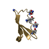
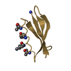
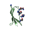
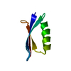
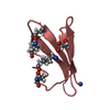
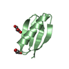
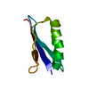
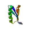
| Title | Nitroxide Spin Labels in Protein GB1: E15 Mutant | ||||||
 Components Components | Immunoglobulin G-binding protein G | ||||||
 Keywords Keywords | IMMUNE SYSTEM / Bacterial Proteins / Crystallization / Electron Spin Resonance Spectroscopy | ||||||
| Function / homology |  Function and homology information Function and homology information | ||||||
| Biological species |  Streptococcus sp. group G (bacteria) Streptococcus sp. group G (bacteria) | ||||||
| Method |  X-RAY DIFFRACTION / MOLECULAR REPLACEMENT / Resolution: 2.2 Å X-RAY DIFFRACTION / MOLECULAR REPLACEMENT / Resolution: 2.2 Å | ||||||
 Authors Authors | Cunningham, T.C. / Horne, W.S. / Saxena, S. | ||||||
 Citation Citation | Journal: Protein Sci. / Year: 2016 Title: Rotameric preferences of a protein spin label at edge-strand beta-sheet sites. Authors: Cunningham, T.F. / Pornsuwan, S. / Horne, W.S. / Saxena, S. | ||||||
| History |
|
- Structure visualization
| Structure viewer | Molecule: MolmilJmol/JSmol |
|---|
- Downloads & links
-Download
| PDBx/mmCIF format | 5bmg.cif.gz | 103.2 KB | Display | PDBx/mmCIF format |
|---|---|---|---|---|
| PDB format | pdb5bmg.ent.gz | 81.3 KB | Display | PDB format |
| PDBx/mmJSON format | 5bmg.json.gz | Tree view | PDBx/mmJSON format | |
| Others |  Other downloads Other downloads |
-Validation report
| Arichive directory | https://data.pdbj.org/pub/pdb/validation_reports/bm/5bmgftp://data.pdbj.org/pub/pdb/validation_reports/bm/5bmg | HTTPS FTP |
|---|
-Related structure data
| Related structure data | 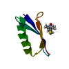 5bmhC 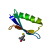 5bmiC 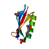 2qmtS C: citing same article ( S: Starting model for refinement |
|---|---|
| Similar structure data |
-Links
 PDBj
PDBj


- Assembly


-Components
| #1: Antibody | Mass: 6202.838 Da / Num. of mol.: 8 / Fragment: UNP residues 304-357 / Mutation: E15C Source method: isolated from a genetically manipulated source Source: (gene. exp.) Streptococcus sp. group G (bacteria) / Gene: spg / Production host: #2: Chemical | ChemComp-MTN /   Mass: 264.385 Da / Num. of mol.: 7 Mass: 264.385 Da / Num. of mol.: 7Source method: isolated from a genetically manipulated source Formula: C10H18NO3S2 #3: Chemical |   Mass: 122.143 Da / Num. of mol.: 2 / Source method: obtained synthetically / Formula: C4H12NO3 / Comment: pH buffer*YM Mass: 122.143 Da / Num. of mol.: 2 / Source method: obtained synthetically / Formula: C4H12NO3 / Comment: pH buffer*YM#4: Water | ChemComp-HOH / |  Mass: 18.015 Da / Num. of mol.: 161 / Source method: isolated from a natural source / Formula: H2O Mass: 18.015 Da / Num. of mol.: 161 / Source method: isolated from a natural source / Formula: H2OHas protein modification | Y | |
|---|
-Experimental details
-Experiment
| Experiment | Method: X-RAY DIFFRACTION / Number of used crystals: 1 |
|---|
- Sample preparation
| Crystal | Density Matthews: 2.12 Å3/Da / Density % sol: 41.89 % |
|---|---|
| Crystal grow | Temperature: 298 K / Method: vapor diffusion, hanging drop / pH: 4.5 Details: 0.1 M magnesium chloride, 0.1 M Tris pH 4.5, 20% w/v PEG 4000 |
-Data collection
| Diffraction | Mean temperature: 100 K |
|---|---|
| Diffraction source | Source: ROTATING ANODE / Type: RIGAKU FR-E SUPERBRIGHT / Wavelength: 1.5418 Å |
| Detector | Type: RIGAKU SATURN 944 / Detector: CCD / Date: Feb 19, 2013 |
| Radiation | Protocol: SINGLE WAVELENGTH / Monochromatic (M) / Laue (L): M / Scattering type: x-ray |
| Radiation wavelength | Wavelength: 1.5418 Å / Relative weight: 1 |
| Reflection | Resolution: 2.2→27.11 Å / Num. obs: 21244 / % possible obs: 97.1 % / Redundancy: 3.2 % / Rmerge(I) obs: 0.084 / Net I/σ(I): 9.1 |
| Reflection shell | Resolution: 2.2→2.28 Å / Redundancy: 2.5 % / Rmerge(I) obs: 0.22 / Mean I/σ(I) obs: 3.7 / % possible all: 90.9 |
- Processing
| Software |
| |||||||||||||||||||||||||||||||||||||||||||||||||||||||||||||||
|---|---|---|---|---|---|---|---|---|---|---|---|---|---|---|---|---|---|---|---|---|---|---|---|---|---|---|---|---|---|---|---|---|---|---|---|---|---|---|---|---|---|---|---|---|---|---|---|---|---|---|---|---|---|---|---|---|---|---|---|---|---|---|---|---|
| Refinement | Method to determine structure: MOLECULAR REPLACEMENT Starting model: 2QMT Resolution: 2.2→27.075 Å / Cross valid method: FREE R-VALUE / σ(F): 1.45 / Phase error: 35.09 / Stereochemistry target values: TWIN_LSQ_F
| |||||||||||||||||||||||||||||||||||||||||||||||||||||||||||||||
| Solvent computation | Shrinkage radii: 0.9 Å / VDW probe radii: 1.11 Å / Solvent model: FLAT BULK SOLVENT MODEL | |||||||||||||||||||||||||||||||||||||||||||||||||||||||||||||||
| Refinement step | Cycle: LAST / Resolution: 2.2→27.075 Å
| |||||||||||||||||||||||||||||||||||||||||||||||||||||||||||||||
| Refine LS restraints |
| |||||||||||||||||||||||||||||||||||||||||||||||||||||||||||||||
| LS refinement shell |
|
