 Movie
Movie Controller


+ Open data

- Basic information
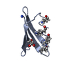
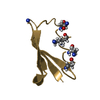
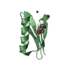
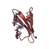
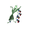
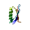
| Entry | Database: PDB / ID: 3gb1 | ||||||
|---|---|---|---|---|---|---|---|
| Title | STRUCTURES OF B1 DOMAIN OF STREPTOCOCCAL PROTEIN G | ||||||
 Components Components | PROTEIN (B1 DOMAIN OF STREPTOCOCCAL PROTEIN G) | ||||||
 Keywords Keywords | IMMUNOGLOBULIN BINDING PROTEIN | ||||||
| Function / homology |  Function and homology information Function and homology information | ||||||
| Biological species |  Streptococcus sp. 'group G' (bacteria) Streptococcus sp. 'group G' (bacteria) | ||||||
| Method | SOLUTION NMR / simulated annealing | ||||||
 Authors Authors | Clore, G.M. | ||||||
 Citation Citation | Journal: J.Am.Chem.Soc. / Year: 1999 Title: Improving the Packing and Accuracy of NMR Structures with a Pseudopotential for the Radius of Gyration Authors: Juszewski, K. / Gronenborn, A.M. / Clore, G.M. #1: Journal: J.Am.Chem.Soc. / Year: 1998Title: Measurement of Residual Dipolar Couplings of Macromolecules Aligned in the Nematic Phase of a Colloidal Suspension of Rod-Shaped Viruses. Authors: Clore, G.M. / Starich, M.R. / Gronenborn, A.M. #2: Journal: Science / Year: 1991Title: A novel, highly stable fold of the immunoglobulin binding domain of streptococcal protein G. Authors: Gronenborn, A.M. / Filpula, D.R. / Essig, N.Z. / Achari, A. / Whitlow, M. / Wingfield, P.T. / Clore, G.M. | ||||||
| History |
|
- Structure visualization
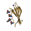
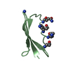
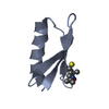
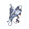
| Structure viewer | Molecule: MolmilJmol/JSmol |
|---|
- Downloads & links
-Download
| PDBx/mmCIF format | 3gb1.cif.gz | 569.2 KB | Display | PDBx/mmCIF format |
|---|---|---|---|---|
| PDB format | pdb3gb1.ent.gz | 484.2 KB | Display | PDB format |
| PDBx/mmJSON format | 3gb1.json.gz | Tree view | PDBx/mmJSON format | |
| Others |  Other downloads Other downloads |
-Validation report
| Arichive directory | https://data.pdbj.org/pub/pdb/validation_reports/gb/3gb1ftp://data.pdbj.org/pub/pdb/validation_reports/gb/3gb1 | HTTPS FTP |
|---|
-Related structure data
| Similar structure data |
|---|
-Links
 PDBj
PDBj- Assembly
| Deposited unit | 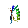
| |||||||||
|---|---|---|---|---|---|---|---|---|---|---|
| 1 |
| |||||||||
| NMR ensembles |
|
-Components
| #1: Protein | Mass: 6201.784 Da / Num. of mol.: 1 / Fragment: B1 DOMAIN Source method: isolated from a genetically manipulated source Source: (gene. exp.) Streptococcus sp. 'group G' (bacteria) / Production host: |
|---|
-Experimental details
-Experiment
| Experiment | Method: SOLUTION NMR | ||||||||||||
|---|---|---|---|---|---|---|---|---|---|---|---|---|---|
| NMR experiment |
|
- Sample preparation
| Sample conditions | Pressure: AMBIENT / Temperature: 298.00 K |
|---|---|
| Crystal grow | *PLUS Method: other / Details: NMR |
-NMR measurement
| NMR spectrometer | Type: Bruker AM600 / Manufacturer: Bruker / Model: AM600 / Field strength: 600 MHz |
|---|
- Processing
| NMR software |
| ||||||||||||
|---|---|---|---|---|---|---|---|---|---|---|---|---|---|
| Refinement | Method: simulated annealing / Software ordinal: 1 Details: INCORPORATES RADIUS OF GYRATION RESTRAINT AND DIPOLAR COUPLINGS A TOTAL OF 31 SIMULATED ANNEALING STRUCTURES WERE CALCULATED THE COORDINATES OF THE RESTRAINED MINIMIZED STRUCTURE ARE LISTED ...Details: INCORPORATES RADIUS OF GYRATION RESTRAINT AND DIPOLAR COUPLINGS A TOTAL OF 31 SIMULATED ANNEALING STRUCTURES WERE CALCULATED THE COORDINATES OF THE RESTRAINED MINIMIZED STRUCTURE ARE LISTED FIRST. THIS WAS OBTAINED BY AVERAGING THE COORDINATES OF THE INDIVIDUAL STRUCTURES AND SUBJECTING THE RESULTING COORDINATES TO RESTRAINED MINIMIZATION. IN THE CASE OF THE RESTRAINED MINIMIZED MEAN STRUCTURE THE QUANTITY PRESENTED IN THE B VALUE FIELD (COLUMNS 61 - 66 OF THE ATOM AND HETATM RECORDS BELOW) REPRESENTS THE ATOMIC RMS DEVIATION OF THE INDIVIDUAL STRUCTURES ABOUT THE MEAN COORDINATE POSITIONS. FOR THE INDIVIDUAL SIMULATED ANNEALING STRUCTURES THE NUMBERS IN THE B-FACTOR C NO MEANING ALL THE INTERPROTON DISTANCE, TORSION ANGLE RESTRAINTS 3G AND DIPOLAR COUPLING RESTRAINTS ARE AVAILABLE FROM THE PROTEIN DATA BANK AS A SEPARATE ENTRY. (RMR3GB1) TERMS IN TARGET FUNCTION USED FOR SIMULATED ANNEALING: NOE (SUM AVERAGING) AND TORSION ANGLE RESTRAINTS 3JHNALPHA COUPLING CONSTANT RESTRAINTS (GARRETT ET AL J. MAGN. RESON. B104, 99-103 (1994). DIPOLAR COUPLING RESTRAINTS USING TWO ALIGNMENT TENSO (IN TMV AND IN BICELLES) TERM FOR THE RADIUS OF GYRATION (KUSZEWSKI J, GRONENB CLORE, GM J AM CHEM SOC 121, 2337-2338 (1999)) TORSION ANGLE DATABASE POTENTIAL (KUSZEWSKI J, GRONEN CLORE GM. PROTEIN SCI 5, 1067-1080 (1996); J. MAGN 125, 171-177 (1997). COVALENT GEOMETRY RESTRAINTS (BONDS, ANGLES, IMPROPER QUARTIC VAN DER WAALS REPULSION TERM (NILGES. M, GRONENBORN, A.M., BRUNGER, A.T., CLORE, G.M. (1988) PROTEIN ENG. 2, 27-38). RESTRAINTS: NOES: 138 SEQUENTIAL, 133 MEDIUM, 279 LONG RANGE IN 185 INTRARESIDUE TORSION ANGLES: 145 3JHNALPHA COUPLINGS: 53 DIPOLAR COUPLINGS: 152 IN TMV AND 148 IN BICELLES (NH, N-C AND HN-C) THE B-FACTOR COLUMN GIVES THE AVERAGE RMS OF THE 31 SIMULATED AN STRUCTURES ABOUT THE MEAN COORDINATE POSITIONS FILENAME=G_TMV_BICE_RGYR_AVE.MIN ============================================================ BONDS,ANGLES,IMPROPERS,CDIH,NOE,COUP 162664E-03,0.47617,0.436105,0,1.84255E-02,0.534352 ============================================================ | ||||||||||||
| NMR ensemble | Conformer selection criteria: RESTRAINED MINIMIZATION / Conformers calculated total number: 32 / Conformers submitted total number: 32 |
