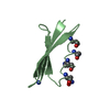
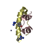
Mass: 6201.784 Da / Num. of mol.: 1 Source method: isolated from a genetically manipulated source Source: (gene. exp.) Streptococcus sp. 'group G' (bacteria) / References: UniProt: P06654
-
Experimental details
-
Experiment
Experiment
Method: SOLUTION NMR
-
Sample preparation
Crystal grow
*PLUS
Method: other / Details: NMR
-
Processing
Software
Name
Classification
X-PLOR
modelbuilding
X-PLOR
refinement
X-PLOR
phasing
NMR software
Name
Developer
Classification
DISGEO
HAVEL,WUTHRICH
refinement
X-PLOR
BRUNGER
refinement
Refinement
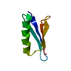
Software ordinal: 1 Details: DETAILS OF THE STRUCTURE DETERMINATION AND ALL STRUCTURAL STATISTICS ARE GIVEN IN THE PAPER CITED ON THE JRNL RECORDS ABOVE (I.E. AGREEMENT WITH EXPERIMENTAL RESTRAINTS, DEVIATIONS FROM ...Details: DETAILS OF THE STRUCTURE DETERMINATION AND ALL STRUCTURAL STATISTICS ARE GIVEN IN THE PAPER CITED ON THE JRNL RECORDS ABOVE (I.E. AGREEMENT WITH EXPERIMENTAL RESTRAINTS, DEVIATIONS FROM IDEALITY FOR BOND LENGTHS, ANGLES, PLANES AND CHIRALITY, NON-BONDED CONTACTS, ATOMIC RMS DIFFERENCES BETWEEN THE CALCULATED STRUCTURES). THE STRUCTURES ARE BASED ON 854 INTERPROTON DISTANCE RESTRAINTS DERIVED FROM NOE MEASUREMENTS; 60 HYDROGEN-BONDING DISTANCE RESTRAINTS FOR 30 HYDROGEN-BONDS IDENTIFIED ON THE BASIS OF THE NOE AND AMIDE PROTON EXCHANGE DATA, AS WELL AS THE INITIAL STRUCTURE CALCULATIONS; AND 54 PHI AND 51 PSI BACKBONE TORSION ANGLE RESTRAINTS AND 39 CHI1 SIDE CHAIN TORSION ANGLE RESTRAINTS DERIVED FROM COUPLING CONSTANTS AND NOE DATA. THE LATTER ARE OBTAINED USING THE CONFORMATIONAL GRID SEARCH PROGRAM STEREOSEARCH [NILGES, M., CLORE, G.M. & GRONENBORN, A.M. (1990) BIOPOLYMERS 29, 813-822. THE METHOD USED TO DETERMINE THE STRUCTURES IS THE HYBRID METRIC MATRIX DISTANCE GEOMETRY-DYNAMICAL SIMULATED ANNEALING METHOD [NILGES, M., CLORE, G.M. & GRONENBORN, A.M. FEBS LETT. 229, 317-324 (1988)]. A TOTAL OF 60 STRUCTURES WERE CALCULATED. THE COORDINATES OF THE RESTRAINED MINIMIZED STRUCTURE ARE PRESENTED IN PROTEIN DATA BANK ENTRY 2GB1. THIS WAS OBTAINED BY AVERAGING THE COORDINATES OF THE INDIVIDUAL STRUCTURES AND SUBJECTING THE RESULTING COORDINATES TO RESTRAINED MINIMIZATION. THE 60 STRUCTURES ARE PRESENTED IN PROTEIN DATA BANK ENTRY 1GB1. THE QUANTITY PRESENTED IN THE B VALUE FIELD (COLUMNS 61 - 66 OF THE ATOM AND HETATM RECORDS BELOW) REPRESENTS THE ATOMIC RMS DEVIATION OF THE INDIVIDUAL STRUCTURES ABOUT THE MEAN COORDINATE POSITIONS.
NMR ensemble
Conformers submitted total number: 1
+
About Yorodumi
-
News
-
Feb 9, 2022. New format data for meta-information of EMDB entries
New format data for meta-information of EMDB entries
Version 3 of the EMDB header file is now the official format.
The previous official version 1.9 will be removed from the archive.
In the structure databanks used in Yorodumi, some data are registered as the other names, "COVID-19 virus" and "2019-nCoV". Here are the details of the virus and the list of structure data.
Jan 31, 2019. EMDB accession codes are about to change! (news from PDBe EMDB page)
EMDB accession codes are about to change! (news from PDBe EMDB page)
The allocation of 4 digits for EMDB accession codes will soon come to an end. Whilst these codes will remain in use, new EMDB accession codes will include an additional digit and will expand incrementally as the available range of codes is exhausted. The current 4-digit format prefixed with “EMD-” (i.e. EMD-XXXX) will advance to a 5-digit format (i.e. EMD-XXXXX), and so on. It is currently estimated that the 4-digit codes will be depleted around Spring 2019, at which point the 5-digit format will come into force.
The EM Navigator/Yorodumi systems omit the EMD- prefix.
Related info.:Q: What is EMD? / ID/Accession-code notation in Yorodumi/EM Navigator
Yorodumi is a browser for structure data from EMDB, PDB, SASBDB, etc.
This page is also the successor to EM Navigator detail page, and also detail information page/front-end page for Omokage search.
The word "yorodu" (or yorozu) is an old Japanese word meaning "ten thousand". "mi" (miru) is to see.
Related info.:EMDB / PDB / SASBDB / Comparison of 3 databanks / Yorodumi Search / Aug 31, 2016. New EM Navigator & Yorodumi / Yorodumi Papers / Jmol/JSmol / Function and homology information / Changes in new EM Navigator and Yorodumi
 Movie
Movie Controller

 Yorodumi
 Open data
 Basic information
 Components
 Keywords
 Function and homology information
 Streptococcus sp. 'group G' (bacteria)
 Authors
 Citation
 Structure visualization
 Downloads & links
 Other downloads
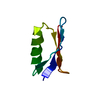


 PDBj
PDBj Assembly
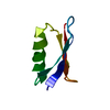
 Sample preparation
 Processing
 X-PLOR