 Movie
Movie Controller

[English] 日本語
 Yorodumi
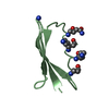
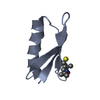
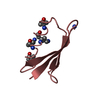
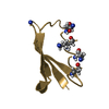
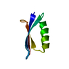
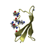
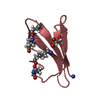
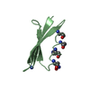
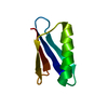
- PDB-6cpz: Selenomethionine mutant (I6Sem) of protein GB1 examined by X-ray ... -
+ Open data

- Basic information
| Entry | Database: PDB / ID: 6cpz | ||||||||||||
|---|---|---|---|---|---|---|---|---|---|---|---|---|---|
| Title | Selenomethionine mutant (I6Sem) of protein GB1 examined by X-ray diffraction | ||||||||||||
 Components Components | Immunoglobulin G-binding protein G | ||||||||||||
 Keywords Keywords | IMMUNE SYSTEM / Immunoglobulin G-binding protein domain B1 | ||||||||||||
| Function / homology |  Function and homology information Function and homology information | ||||||||||||
| Biological species |  Streptococcus sp. group G (bacteria) Streptococcus sp. group G (bacteria) | ||||||||||||
| Method |  X-RAY DIFFRACTION / SYNCHROTRON / MOLECULAR REPLACEMENT / Resolution: 1.12 Å X-RAY DIFFRACTION / SYNCHROTRON / MOLECULAR REPLACEMENT / Resolution: 1.12 Å | ||||||||||||
 Authors Authors | Chen, Q. / Rozovsky, S. | ||||||||||||
| Funding support |  United States, 3items United States, 3items
| ||||||||||||
 Citation Citation | Journal: J.Phys.Chem.B / Year: 2020 Title: 77Se NMR Probes the Protein Environment of Selenomethionine. Authors: Chen, Q. / Xu, S. / Lu, X. / Boeri, M.V. / Pepelyayeva, Y. / Diaz, E.L. / Soni, S.D. / Allaire, M. / Forstner, M.B. / Bahnson, B.J. / Rozovsky, S. | ||||||||||||
| History |
|
- Structure visualization
| Structure viewer | Molecule: MolmilJmol/JSmol |
|---|
- Downloads & links
-Download
| PDBx/mmCIF format | 6cpz.cif.gz | 83.9 KB | Display | PDBx/mmCIF format |
|---|---|---|---|---|
| PDB format | pdb6cpz.ent.gz | 65.2 KB | Display | PDB format |
| PDBx/mmJSON format | 6cpz.json.gz | Tree view | PDBx/mmJSON format | |
| Others |  Other downloads Other downloads |
-Validation report
| Arichive directory | https://data.pdbj.org/pub/pdb/validation_reports/cp/6cpzftp://data.pdbj.org/pub/pdb/validation_reports/cp/6cpz | HTTPS FTP |
|---|
-Related structure data
| Related structure data | 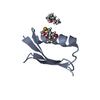 6c9oC 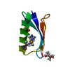 6cheC 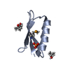 6cneC 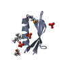 6cteC 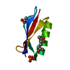 2qmtS C: citing same article ( S: Starting model for refinement |
|---|---|
| Similar structure data |

-Links
 PDBj
PDBj

- Assembly
| Deposited unit | 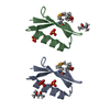
| ||||||||
|---|---|---|---|---|---|---|---|---|---|
| 1 | 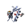
| ||||||||
| 2 | 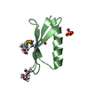
| ||||||||
| Unit cell |
|
-Components
| #1: Antibody | Mass: 6219.598 Da / Num. of mol.: 2 / Mutation: I6Sem Source method: isolated from a genetically manipulated source Source: (gene. exp.) Streptococcus sp. group G (bacteria) / Gene: spg / Cell line (production host): BL21(DE3) / Production host: #2: Chemical |   Mass: 118.174 Da / Num. of mol.: 2 / Source method: obtained synthetically / Formula: C6H14O2 / Comment: precipitant*YM Mass: 118.174 Da / Num. of mol.: 2 / Source method: obtained synthetically / Formula: C6H14O2 / Comment: precipitant*YM#3: Chemical |   Mass: 118.174 Da / Num. of mol.: 2 / Source method: obtained synthetically / Formula: C6H14O2 / Comment: precipitant*YM Mass: 118.174 Da / Num. of mol.: 2 / Source method: obtained synthetically / Formula: C6H14O2 / Comment: precipitant*YM#4: Chemical | ChemComp-PO4 /   Mass: 94.971 Da / Num. of mol.: 4 / Source method: obtained synthetically / Formula: PO4 Mass: 94.971 Da / Num. of mol.: 4 / Source method: obtained synthetically / Formula: PO4#5: Water | ChemComp-HOH / |  Mass: 18.015 Da / Num. of mol.: 179 / Source method: isolated from a natural source / Formula: H2O Mass: 18.015 Da / Num. of mol.: 179 / Source method: isolated from a natural source / Formula: H2OHas protein modification | Y | |
|---|
-Experimental details
-Experiment
| Experiment | Method: X-RAY DIFFRACTION / Number of used crystals: 1 |
|---|
- Sample preparation
| Crystal | Density Matthews: 1.99 Å3/Da / Density % sol: 38.32 % |
|---|---|
| Crystal grow | Temperature: 283.15 K / Method: vapor diffusion, hanging drop / pH: 4.7 Details: 48% MPD 20% IPA 25 mM sodium acetate buffer pH 4.7 20 mg/ml protein in 25 mM sodium acetate buffer pH 5.5 and 2 mM TCEP PH range: 4.5-4.9 |
-Data collection
| Diffraction | Mean temperature: 100 K |
|---|---|
| Diffraction source | Source: SYNCHROTRON / Site: ALS / Beamline: 5.0.1 / Wavelength: 0.9795 Å |
| Detector | Type: DECTRIS PILATUS3 6M / Detector: PIXEL / Date: Aug 17, 2017 |
| Radiation | Monochromator: Asymetric curved crystal / Protocol: SINGLE WAVELENGTH / Monochromatic (M) / Laue (L): M / Scattering type: x-ray |
| Radiation wavelength | Wavelength: 0.9795 Å / Relative weight: 1 |
| Reflection | Resolution: 1.12→27.571 Å / Num. obs: 69779 / % possible obs: 97.43 % / Redundancy: 2 % / Biso Wilson estimate: 7.95 Å2 / CC1/2: 0.999 / Rmerge(I) obs: 0.01784 / Rpim(I) all: 0.01784 / Rrim(I) all: 0.02524 / Net I/σ(I): 12.98 |
| Reflection shell | Resolution: 1.12→1.16 Å |
- Processing
| Software |
| ||||||||||||||||||||||||||||||||||||||||||||||||||||||||||||||||||||||||||||||||||||||||||||||||||||||||||||||||||||||||||||||||||||||||||||||||||||||||||||||||||||||||||||||||||||||||||||||||||||
|---|---|---|---|---|---|---|---|---|---|---|---|---|---|---|---|---|---|---|---|---|---|---|---|---|---|---|---|---|---|---|---|---|---|---|---|---|---|---|---|---|---|---|---|---|---|---|---|---|---|---|---|---|---|---|---|---|---|---|---|---|---|---|---|---|---|---|---|---|---|---|---|---|---|---|---|---|---|---|---|---|---|---|---|---|---|---|---|---|---|---|---|---|---|---|---|---|---|---|---|---|---|---|---|---|---|---|---|---|---|---|---|---|---|---|---|---|---|---|---|---|---|---|---|---|---|---|---|---|---|---|---|---|---|---|---|---|---|---|---|---|---|---|---|---|---|---|---|---|---|---|---|---|---|---|---|---|---|---|---|---|---|---|---|---|---|---|---|---|---|---|---|---|---|---|---|---|---|---|---|---|---|---|---|---|---|---|---|---|---|---|---|---|---|---|---|---|---|
| Refinement | Method to determine structure: MOLECULAR REPLACEMENT Starting model: 2QMT Resolution: 1.12→27.571 Å / SU ML: 0.07 / Cross valid method: FREE R-VALUE / σ(F): 1.36 / Phase error: 18.1 / Stereochemistry target values: ML
| ||||||||||||||||||||||||||||||||||||||||||||||||||||||||||||||||||||||||||||||||||||||||||||||||||||||||||||||||||||||||||||||||||||||||||||||||||||||||||||||||||||||||||||||||||||||||||||||||||||
| Solvent computation | Shrinkage radii: 0.9 Å / VDW probe radii: 1.11 Å / Solvent model: FLAT BULK SOLVENT MODEL | ||||||||||||||||||||||||||||||||||||||||||||||||||||||||||||||||||||||||||||||||||||||||||||||||||||||||||||||||||||||||||||||||||||||||||||||||||||||||||||||||||||||||||||||||||||||||||||||||||||
| Refinement step | Cycle: LAST / Resolution: 1.12→27.571 Å
| ||||||||||||||||||||||||||||||||||||||||||||||||||||||||||||||||||||||||||||||||||||||||||||||||||||||||||||||||||||||||||||||||||||||||||||||||||||||||||||||||||||||||||||||||||||||||||||||||||||
| Refine LS restraints |
| ||||||||||||||||||||||||||||||||||||||||||||||||||||||||||||||||||||||||||||||||||||||||||||||||||||||||||||||||||||||||||||||||||||||||||||||||||||||||||||||||||||||||||||||||||||||||||||||||||||
| LS refinement shell |
|
