 Movie
Movie Controller


+ Open data

- Basic information
| Entry | Database: PDB / ID: 9ins | ||||||
|---|---|---|---|---|---|---|---|
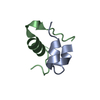
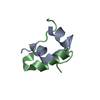
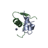
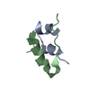
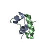
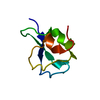
| Title | MONOVALENT CATION BINDING IN CUBIC INSULIN CRYSTALS | ||||||
 Components Components |
| ||||||
 Keywords Keywords | HORMONE | ||||||
| Function / homology |  Function and homology information Function and homology informationpositive regulation of lipoprotein lipase activity / Insulin processing / IRS activation / Signal attenuation / Insulin receptor signalling cascade / Signaling by Insulin receptor / Synthesis, secretion, and deacylation of Ghrelin / PI5P, PP2A and IER3 Regulate PI3K/AKT Signaling / Insulin receptor recycling / response to L-arginine ...positive regulation of lipoprotein lipase activity / Insulin processing / IRS activation / Signal attenuation / Insulin receptor signalling cascade / Signaling by Insulin receptor / Synthesis, secretion, and deacylation of Ghrelin / PI5P, PP2A and IER3 Regulate PI3K/AKT Signaling / Insulin receptor recycling / response to L-arginine / lactate biosynthetic process / positive regulation of glucose metabolic process / positive regulation of fatty acid biosynthetic process / glycoprotein biosynthetic process / lipoprotein biosynthetic process / COPI-mediated anterograde transport / negative regulation of glycogen catabolic process / : / negative regulation of fatty acid metabolic process / negative regulation of feeding behavior / lipid biosynthetic process / negative regulation of acute inflammatory response / positive regulation of respiratory burst / alpha-beta T cell activation / TORC1 signaling / negative regulation of protein secretion / positive regulation of dendritic spine maintenance / negative regulation of gluconeogenesis / fatty acid homeostasis / positive regulation of glycogen biosynthetic process / positive regulation of insulin receptor signaling pathway / negative regulation of respiratory burst involved in inflammatory response / negative regulation of lipid catabolic process / negative regulation of ubiquitin-dependent protein catabolic process / nitric oxide-cGMP-mediated signaling / regulation of protein localization to plasma membrane / negative regulation of reactive oxygen species biosynthetic process / insulin-like growth factor receptor binding / neuron projection maintenance / positive regulation of mitotic nuclear division / positive regulation of DNA replication / positive regulation of glycolytic process / positive regulation of cytokine production / acute-phase response / positive regulation of D-glucose import across plasma membrane / insulin receptor binding / positive regulation of protein secretion / positive regulation of translation / wound healing / hormone activity / positive regulation of neuron projection development / negative regulation of protein catabolic process / positive regulation of protein localization to nucleus / glucose metabolic process / vasodilation / insulin receptor signaling pathway / glucose homeostasis / protease binding / positive regulation of MAPK cascade / positive regulation of canonical NF-kappaB signal transduction / positive regulation of phosphatidylinositol 3-kinase/protein kinase B signal transduction / positive regulation of cell migration / G protein-coupled receptor signaling pathway / negative regulation of gene expression / positive regulation of cell population proliferation / : / identical protein binding Similarity search - Function | ||||||
| Biological species |  | ||||||
| Method |  X-RAY DIFFRACTION / Resolution: 1.7 Å X-RAY DIFFRACTION / Resolution: 1.7 Å | ||||||
 Authors Authors | Badger, J. / Dodson, G.G. | ||||||
 Citation Citation | Journal: Biophys.J. / Year: 1992 Title: Monovalent cation binding to cubic insulin crystals Authors: Gursky, O. / Li, Y. / Badger, J. / Caspar, D.L.D. #1: Journal: Acta Crystallogr.,Sect.B / Year: 1991Title: Structure of the Pig Insulin Dimer in the Cubic Crystal Authors: Badger, J. / Harris, M.R. / Reynolds, C.D. / Evans, A.C. / Dodson, E.J. / Dodson, G.G. / North, A.C.T. #2: Journal: Proc.Natl.Acad.Sci.USA / Year: 1991Title: Water Structure in Cubic Insulin Crystals Authors: Badger, J. / Caspar, D.L.D. #3: Journal: J.Mol.Biol. / Year: 1978Title: Zinc-Free Cubic Pig Insulin: Crystallization and Structure Determination Authors: Dodson, E.J. / Dodson, G.G. / Lewitova, A. / Sabesan, M. | ||||||
| History |
|
- Structure visualization
| Structure viewer | Molecule: MolmilJmol/JSmol |
|---|
- Downloads & links
-Download
| PDBx/mmCIF format | 9ins.cif.gz | 23.7 KB | Display | PDBx/mmCIF format |
|---|---|---|---|---|
| PDB format | pdb9ins.ent.gz | 15.8 KB | Display | PDB format |
| PDBx/mmJSON format | 9ins.json.gz | Tree view | PDBx/mmJSON format | |
| Others |  Other downloads Other downloads |
-Validation report
| Arichive directory | https://data.pdbj.org/pub/pdb/validation_reports/in/9insftp://data.pdbj.org/pub/pdb/validation_reports/in/9ins | HTTPS FTP |
|---|
-Related structure data
| Similar structure data |
|---|


-Links
 PDBj
PDBj


- Assembly
| Deposited unit | 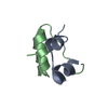
| |||||||||
|---|---|---|---|---|---|---|---|---|---|---|
| 1 |
| |||||||||
| 2 | 
| |||||||||
| Unit cell |
| |||||||||
| Atom site foot note | 1: WATER 24 IS PROBABLY A PARTIALLY OCCUPIED SODIUM ION. ANOTHER SODIUM SITE WITH VERY LOW OCCUPANCY OVERLAPS THE MAJOR CONFORMATION OF HIS B10 (SEE REF 3). 2: SEE REMARK 5. | |||||||||
| Components on special symmetry positions |
|
-Components
| #1: Protein/peptide | Mass: 2383.698 Da / Num. of mol.: 1 Source method: isolated from a genetically manipulated source Source: (gene. exp.) |
|---|---|
| #2: Protein/peptide | Mass: 3403.927 Da / Num. of mol.: 1 Source method: isolated from a genetically manipulated source Source: (gene. exp.) |
| #3: Water | ChemComp-HOH /  Mass: 18.015 Da / Num. of mol.: 81 / Source method: isolated from a natural source / Formula: H2O Mass: 18.015 Da / Num. of mol.: 81 / Source method: isolated from a natural source / Formula: H2O |
| Has protein modification | Y |
-Experimental details
-Experiment
| Experiment | Method: X-RAY DIFFRACTION |
|---|
- Sample preparation
| Crystal | Density Matthews: 3.54 Å3/Da / Density % sol: 65.21 % | ||||||||||||||||||||||||||||||||||||||||||
|---|---|---|---|---|---|---|---|---|---|---|---|---|---|---|---|---|---|---|---|---|---|---|---|---|---|---|---|---|---|---|---|---|---|---|---|---|---|---|---|---|---|---|---|
| Crystal grow | *PLUS Method: microdialysis | ||||||||||||||||||||||||||||||||||||||||||
| Components of the solutions | *PLUS
|
- Processing
| Software | Name: PROLSQ / Classification: refinement | ||||||||||||||||||||||||||||||||||||||||||||||||||||||||||||||||||||||||||||||||||||
|---|---|---|---|---|---|---|---|---|---|---|---|---|---|---|---|---|---|---|---|---|---|---|---|---|---|---|---|---|---|---|---|---|---|---|---|---|---|---|---|---|---|---|---|---|---|---|---|---|---|---|---|---|---|---|---|---|---|---|---|---|---|---|---|---|---|---|---|---|---|---|---|---|---|---|---|---|---|---|---|---|---|---|---|---|---|
| Refinement | Resolution: 1.7→10 Å / Rfactor obs: 0.178 / σ(F): 2 Details: BY COMPARISON WITH MULTIPLE DATA SETS FROM BOVINE CUBIC INSULIN COLLECTED IN THE PH RANGE 7 TO 11 (REFERENCE 1 AND UNPUBLISHED RESULTS) IT HAS BEEN POSSIBLE TO RELIABLY IDENTIFY ADDITIONAL ...Details: BY COMPARISON WITH MULTIPLE DATA SETS FROM BOVINE CUBIC INSULIN COLLECTED IN THE PH RANGE 7 TO 11 (REFERENCE 1 AND UNPUBLISHED RESULTS) IT HAS BEEN POSSIBLE TO RELIABLY IDENTIFY ADDITIONAL CONFORMATIONS FOR SEVERAL SIDE CHAINS THAT WERE NOT INCLUDED IN THE PUBLISHED (ACTA CRYSTALLOGR.) REPORT. THESE HAVE BEEN MODELED, THE ORDERED SOLVENT RATIONALIZED AND THE STRUCTURE REFINED FURTHER. THE R-FACTOR AND STEREOCHEMICAL PARAMETERS IN THIS ENTRY ARE FOR THIS NEW MODEL. IN THE ACTA CRYSTALLOGRAPHICA 1991 PUBLICATION (REFERENCE 2 ABOVE) REFERENCE IS MADE TO PROTEIN DATA BANK ENTRY 5INS. THE COORDINATES IN THIS ENTRY SUPERSEDE 5INS. | ||||||||||||||||||||||||||||||||||||||||||||||||||||||||||||||||||||||||||||||||||||
| Refinement step | Cycle: LAST / Resolution: 1.7→10 Å
| ||||||||||||||||||||||||||||||||||||||||||||||||||||||||||||||||||||||||||||||||||||
| Refine LS restraints |
| ||||||||||||||||||||||||||||||||||||||||||||||||||||||||||||||||||||||||||||||||||||
| Refinement | *PLUS Rfactor Rwork: 0.178 | ||||||||||||||||||||||||||||||||||||||||||||||||||||||||||||||||||||||||||||||||||||
| Solvent computation | *PLUS | ||||||||||||||||||||||||||||||||||||||||||||||||||||||||||||||||||||||||||||||||||||
| Displacement parameters | *PLUS |
