 Movie
Movie Controller

[English] 日本語
 Yorodumi
- PDB-5d52: In meso in situ serial X-ray crystallography structure of insulin... -
+ Open data

- Basic information
| Entry | Database: PDB / ID: 5d52 | ||||||
|---|---|---|---|---|---|---|---|
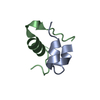
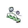
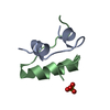
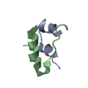
| Title | In meso in situ serial X-ray crystallography structure of insulin at room temperature | ||||||
 Components Components |
| ||||||
 Keywords Keywords | HORMONE | ||||||
| Function / homology |  Function and homology information Function and homology informationpositive regulation of lipoprotein lipase activity / Insulin processing / IRS activation / Signal attenuation / Insulin receptor signalling cascade / Signaling by Insulin receptor / Synthesis, secretion, and deacylation of Ghrelin / PI5P, PP2A and IER3 Regulate PI3K/AKT Signaling / Insulin receptor recycling / response to L-arginine ...positive regulation of lipoprotein lipase activity / Insulin processing / IRS activation / Signal attenuation / Insulin receptor signalling cascade / Signaling by Insulin receptor / Synthesis, secretion, and deacylation of Ghrelin / PI5P, PP2A and IER3 Regulate PI3K/AKT Signaling / Insulin receptor recycling / response to L-arginine / lactate biosynthetic process / positive regulation of glucose metabolic process / positive regulation of fatty acid biosynthetic process / glycoprotein biosynthetic process / lipoprotein biosynthetic process / COPI-mediated anterograde transport / negative regulation of glycogen catabolic process / : / negative regulation of fatty acid metabolic process / negative regulation of feeding behavior / lipid biosynthetic process / negative regulation of acute inflammatory response / positive regulation of respiratory burst / alpha-beta T cell activation / TORC1 signaling / negative regulation of protein secretion / positive regulation of dendritic spine maintenance / negative regulation of gluconeogenesis / fatty acid homeostasis / positive regulation of glycogen biosynthetic process / positive regulation of insulin receptor signaling pathway / negative regulation of respiratory burst involved in inflammatory response / negative regulation of lipid catabolic process / negative regulation of ubiquitin-dependent protein catabolic process / nitric oxide-cGMP-mediated signaling / regulation of protein localization to plasma membrane / negative regulation of reactive oxygen species biosynthetic process / insulin-like growth factor receptor binding / neuron projection maintenance / positive regulation of mitotic nuclear division / positive regulation of DNA replication / positive regulation of glycolytic process / positive regulation of cytokine production / acute-phase response / positive regulation of D-glucose import across plasma membrane / insulin receptor binding / positive regulation of protein secretion / positive regulation of translation / wound healing / hormone activity / positive regulation of neuron projection development / negative regulation of protein catabolic process / positive regulation of protein localization to nucleus / glucose metabolic process / vasodilation / insulin receptor signaling pathway / glucose homeostasis / protease binding / positive regulation of MAPK cascade / positive regulation of canonical NF-kappaB signal transduction / positive regulation of phosphatidylinositol 3-kinase/protein kinase B signal transduction / positive regulation of cell migration / G protein-coupled receptor signaling pathway / negative regulation of gene expression / positive regulation of cell population proliferation / : / identical protein binding Similarity search - Function | ||||||
| Biological species |  | ||||||
| Method |  X-RAY DIFFRACTION / SYNCHROTRON / MOLECULAR REPLACEMENT / Resolution: 1.8 Å X-RAY DIFFRACTION / SYNCHROTRON / MOLECULAR REPLACEMENT / Resolution: 1.8 Å | ||||||
 Authors Authors | Huang, C.-Y. / Olieric, V. / Warshamanage, R. / Diederichs, K. / Wang, M. / Caffrey, M. | ||||||
| Funding support |  Ireland, 1items Ireland, 1items
| ||||||
 Citation Citation | Journal: Acta Crystallogr D Struct Biol / Year: 2016 Title: In meso in situ serial X-ray crystallography of soluble and membrane proteins at cryogenic temperatures. Authors: Huang, C.Y. / Olieric, V. / Ma, P. / Howe, N. / Vogeley, L. / Liu, X. / Warshamanage, R. / Weinert, T. / Panepucci, E. / Kobilka, B. / Diederichs, K. / Wang, M. / Caffrey, M. | ||||||
| History |
|
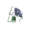
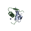
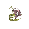
- Structure visualization
| Structure viewer | Molecule: MolmilJmol/JSmol |
|---|
- Downloads & links
-Download
| PDBx/mmCIF format | 5d52.cif.gz | 24.1 KB | Display | PDBx/mmCIF format |
|---|---|---|---|---|
| PDB format | pdb5d52.ent.gz | 14.5 KB | Display | PDB format |
| PDBx/mmJSON format | 5d52.json.gz | Tree view | PDBx/mmJSON format | |
| Others |  Other downloads Other downloads |
-Validation report
| Arichive directory | https://data.pdbj.org/pub/pdb/validation_reports/d5/5d52ftp://data.pdbj.org/pub/pdb/validation_reports/d5/5d52 | HTTPS FTP |
|---|
-Related structure data
| Related structure data | 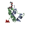 5d53C 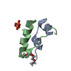 5d54C 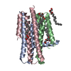 5d56C  5d57C 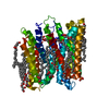 5d58C 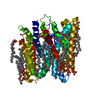 5d59C 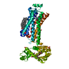 5d5aC  5d5bC 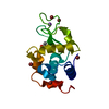 5d5cC 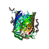 5d5dC 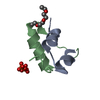 5d5eC 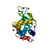 5d5fC 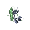 9insS S: Starting model for refinement C: citing same article ( |
|---|---|
| Similar structure data |


-Links
 PDBj
PDBj


- Assembly
| Deposited unit | 
| |||||||||
|---|---|---|---|---|---|---|---|---|---|---|
| 1 |
| |||||||||
| Unit cell |
| |||||||||
| Components on special symmetry positions |
|
-Components
| #1: Protein/peptide | Mass: 2383.698 Da / Num. of mol.: 1 / Source method: isolated from a natural source / Source: (natural) | ||||
|---|---|---|---|---|---|
| #2: Protein/peptide | Mass: 3403.927 Da / Num. of mol.: 1 / Source method: isolated from a natural source / Source: (natural) | ||||
| #3: Chemical |   Mass: 94.971 Da / Num. of mol.: 2 / Source method: obtained synthetically / Formula: PO4 Mass: 94.971 Da / Num. of mol.: 2 / Source method: obtained synthetically / Formula: PO4#4: Water | ChemComp-HOH / |  Mass: 18.015 Da / Num. of mol.: 30 / Source method: isolated from a natural source / Formula: H2O Mass: 18.015 Da / Num. of mol.: 30 / Source method: isolated from a natural source / Formula: H2OHas protein modification | Y | |
-Experimental details
-Experiment
| Experiment | Method: X-RAY DIFFRACTION |
|---|
- Sample preparation
| Crystal | Density Matthews: 3.65 Å3/Da / Density % sol: 66.27 % |
|---|---|
| Crystal grow | Temperature: 293 K / Method: lipidic cubic phase Details: 0.1-0.2 M sodium phosphate, pH 5.5-6.1, and 33-38 %(w/v) PEG400 PH range: pH 5.5-6.1 |
-Data collection
| Diffraction | Mean temperature: 293 K |
|---|---|
| Diffraction source | Source: SYNCHROTRON / Site: SLS  / Beamline: X10SA / Wavelength: 1 Å / Beamline: X10SA / Wavelength: 1 Å |
| Detector | Type: DECTRIS PILATUS 6M-F / Detector: PIXEL / Date: Feb 18, 2015 |
| Radiation | Protocol: SINGLE WAVELENGTH / Monochromatic (M) / Laue (L): M / Scattering type: x-ray |
| Radiation wavelength | Wavelength: 1 Å / Relative weight: 1 |
| Reflection | Resolution: 1.8→50 Å / Num. obs: 7975 / % possible obs: 100 % / Redundancy: 13.2 % / Net I/σ(I): 6.96 |
| Reflection shell | Resolution: 1.8→1.85 Å / Redundancy: 13 % / Mean I/σ(I) obs: 0.88 / % possible all: 100 |
- Processing
| Software |
| ||||||||||||||||||||||||||||
|---|---|---|---|---|---|---|---|---|---|---|---|---|---|---|---|---|---|---|---|---|---|---|---|---|---|---|---|---|---|
| Refinement | Method to determine structure: MOLECULAR REPLACEMENT Starting model: 9ins Resolution: 1.8→39.855 Å / SU ML: 0.25 / Cross valid method: THROUGHOUT / σ(F): 1.36 / Phase error: 23.58 / Stereochemistry target values: ML
| ||||||||||||||||||||||||||||
| Solvent computation | Shrinkage radii: 0.9 Å / VDW probe radii: 1.11 Å / Solvent model: FLAT BULK SOLVENT MODEL | ||||||||||||||||||||||||||||
| Refinement step | Cycle: LAST / Resolution: 1.8→39.855 Å
| ||||||||||||||||||||||||||||
| Refine LS restraints |
| ||||||||||||||||||||||||||||
| LS refinement shell |
|
