- PDB-5lis: Insulin solved by Native SAD from a dataset collected in one second -
+
Open data
ID or keywords:
Loading...
-
Basic information
Entry
Database: PDB / ID: 5lis
Title
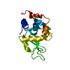
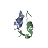
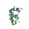
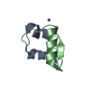
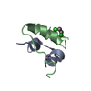
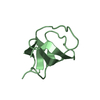
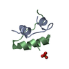
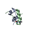
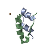
Insulin solved by Native SAD from a dataset collected in one second
Components
(Insulin) x 2
Keywords
HYDROLASE / Insulin
Function / homology
Function and homology information
positive regulation of lipoprotein lipase activity / Insulin processing / IRS activation / Signal attenuation / Insulin receptor signalling cascade / Signaling by Insulin receptor / Synthesis, secretion, and deacylation of Ghrelin / PI5P, PP2A and IER3 Regulate PI3K/AKT Signaling / Insulin receptor recycling / response to L-arginine ...positive regulation of lipoprotein lipase activity / Insulin processing / IRS activation / Signal attenuation / Insulin receptor signalling cascade / Signaling by Insulin receptor / Synthesis, secretion, and deacylation of Ghrelin / PI5P, PP2A and IER3 Regulate PI3K/AKT Signaling / Insulin receptor recycling / response to L-arginine / lactate biosynthetic process / positive regulation of glucose metabolic process / positive regulation of fatty acid biosynthetic process / glycoprotein biosynthetic process / lipoprotein biosynthetic process / COPI-mediated anterograde transport / negative regulation of glycogen catabolic process / : / negative regulation of fatty acid metabolic process / negative regulation of feeding behavior / lipid biosynthetic process / negative regulation of acute inflammatory response / positive regulation of respiratory burst / alpha-beta T cell activation / TORC1 signaling / negative regulation of protein secretion / positive regulation of dendritic spine maintenance / negative regulation of gluconeogenesis / fatty acid homeostasis / positive regulation of glycogen biosynthetic process / positive regulation of insulin receptor signaling pathway / negative regulation of respiratory burst involved in inflammatory response / negative regulation of lipid catabolic process / negative regulation of ubiquitin-dependent protein catabolic process / nitric oxide-cGMP-mediated signaling / regulation of protein localization to plasma membrane / negative regulation of reactive oxygen species biosynthetic process / insulin-like growth factor receptor binding / neuron projection maintenance / positive regulation of mitotic nuclear division / positive regulation of DNA replication / positive regulation of glycolytic process / positive regulation of cytokine production / acute-phase response / positive regulation of D-glucose import across plasma membrane / insulin receptor binding / positive regulation of protein secretion / positive regulation of translation / wound healing / hormone activity / positive regulation of neuron projection development / negative regulation of protein catabolic process / positive regulation of protein localization to nucleus / glucose metabolic process / vasodilation / insulin receptor signaling pathway / glucose homeostasis / protease binding / positive regulation of MAPK cascade / positive regulation of canonical NF-kappaB signal transduction / positive regulation of phosphatidylinositol 3-kinase/protein kinase B signal transduction / positive regulation of cell migration / G protein-coupled receptor signaling pathway / negative regulation of gene expression / positive regulation of cell population proliferation / : / identical protein binding Similarity search - Function
Insulin / Insulin family / Insulin-like / Insulin/IGF/Relaxin family / Insulin / insulin-like growth factor / relaxin family. / Insulin, conserved site / Insulin family signature. / Insulin-like superfamily Similarity search - Domain/homology
Type: DECTRIS EIGER X 16M / Detector: PIXEL / Date: Feb 8, 2015
Radiation
Protocol: SINGLE WAVELENGTH / Monochromatic (M) / Laue (L): M / Scattering type: x-ray
Radiation wavelength
Wavelength: 1.55 Å / Relative weight: 1
Reflection
Resolution: 2.293→38.44 Å / Num. obs: 5420 / % possible obs: 85 % / Redundancy: 5.4 % / Rrim(I) all: 0.027 / Net I/σ(I): 31.14
Reflection shell
Highest resolution: 2.293 Å
-
Processing
Software
Name
Version
Classification
REFMAC
5.8.0155
refinement
XDS
datareduction
XDS
datascaling
CRANK2
phasing
Refinement
Method to determine structure: SAD / Resolution: 2.293→38.44 Å / Cor.coef. Fo:Fc: 0.934 / Cor.coef. Fo:Fc free: 0.908 / SU B: 13.118 / SU ML: 0.148 / Cross valid method: THROUGHOUT / ESU R: 0.331 / ESU R Free: 0.253 / Details: HYDROGENS HAVE BEEN ADDED IN THE RIDING POSITIONS
Rfactor
Num. reflection
% reflection
Selection details
Rfree
0.26186
303
10.1 %
RANDOM
Rwork
0.21697
-
-
-
obs
0.22149
2706
85.48 %
-
Solvent computation
Ion probe radii: 0.8 Å / Shrinkage radii: 0.8 Å / VDW probe radii: 1 Å
 Movie
Movie Controller

 Yorodumi
 Open data
 Basic information
 Components
 Keywords
 Function and homology information
 X-RAY DIFFRACTION /
  Authors
 Citation
 Structure visualization
 Downloads & links
 Other downloads
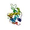


 PDBj
PDBj


 Assembly
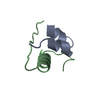
 Mass: 18.015 Da / Num. of mol.: 3 / Source method: isolated from a natural source / Formula: H2O
 Sample preparation
 / Beamline: X10SA / Wavelength: 1.55 Å
 Processing