 Movie
Movie Controller

[English] 日本語
 Yorodumi
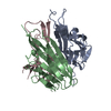
- PDB-5d5d: In meso in situ serial X-ray crystallography structure of AlgE at... -
+ Open data

- Basic information
| Entry | Database: PDB / ID: 5d5d | |||||||||
|---|---|---|---|---|---|---|---|---|---|---|
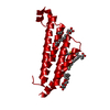
| Title | In meso in situ serial X-ray crystallography structure of AlgE at 100 K | |||||||||
 Components Components | Alginate production protein AlgE | |||||||||
 Keywords Keywords | TRANSPORT PROTEIN / Beta-Barrel Membrane Proteins / AlgE alginate export protein | |||||||||
| Function / homology |  Function and homology information Function and homology information | |||||||||
| Biological species |   Pseudomonas aeruginosa (bacteria) Pseudomonas aeruginosa (bacteria) | |||||||||
| Method |  X-RAY DIFFRACTION / SYNCHROTRON / MOLECULAR REPLACEMENT / Resolution: 2.4 Å X-RAY DIFFRACTION / SYNCHROTRON / MOLECULAR REPLACEMENT / Resolution: 2.4 Å | |||||||||
 Authors Authors | Ma, P. / Huang, C.-Y. / Olieric, V. / Diederichs, K. / Wang, M. / Caffrey, M. | |||||||||
| Funding support |  Ireland, Ireland,  Belgium, 2items Belgium, 2items
| |||||||||
 Citation Citation | Journal: Acta Crystallogr D Struct Biol / Year: 2016 Title: In meso in situ serial X-ray crystallography of soluble and membrane proteins at cryogenic temperatures. Authors: Huang, C.Y. / Olieric, V. / Ma, P. / Howe, N. / Vogeley, L. / Liu, X. / Warshamanage, R. / Weinert, T. / Panepucci, E. / Kobilka, B. / Diederichs, K. / Wang, M. / Caffrey, M. | |||||||||
| History |
|
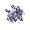
- Structure visualization
| Structure viewer | Molecule: MolmilJmol/JSmol |
|---|
- Downloads & links
-Download
| PDBx/mmCIF format | 5d5d.cif.gz | 105.8 KB | Display | PDBx/mmCIF format |
|---|---|---|---|---|
| PDB format | pdb5d5d.ent.gz | 78.5 KB | Display | PDB format |
| PDBx/mmJSON format | 5d5d.json.gz | Tree view | PDBx/mmJSON format | |
| Others |  Other downloads Other downloads |
-Validation report
| Arichive directory | https://data.pdbj.org/pub/pdb/validation_reports/d5/5d5dftp://data.pdbj.org/pub/pdb/validation_reports/d5/5d5d | HTTPS FTP |
|---|
-Related structure data
| Related structure data |  5d52C  5d53C  5d54C 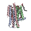 5d56C  5d57C  5d58C  5d59C  5d5aC  5d5bC 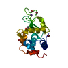 5d5cC  5d5eC 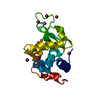 5d5fC  4afkS S: Starting model for refinement C: citing same article ( |
|---|---|
| Similar structure data |


-Links
 PDBj
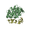
PDBj- Assembly
| Deposited unit | 
| ||||||||
|---|---|---|---|---|---|---|---|---|---|
| 1 |
| ||||||||
| Unit cell |
|
-Components
-Protein , 1 types, 1 molecules A
| #1: Protein | Mass: 54481.355 Da / Num. of mol.: 1 Source method: isolated from a genetically manipulated source Source: (gene. exp.) Pseudomonas aeruginosa (strain ATCC 15692 / PAO1 / 1C / PRS 101 / LMG 12228) (bacteria)Gene: algE, alg76, PA3544 / Production host: |
|---|
-Non-polymers , 7 types, 42 molecules 


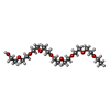


| #2: Chemical |  Mass: 22.990 Da / Num. of mol.: 2 / Source method: obtained synthetically / Formula: Na Mass: 22.990 Da / Num. of mol.: 2 / Source method: obtained synthetically / Formula: Na#3: Chemical | ChemComp-CA / |  Mass: 40.078 Da / Num. of mol.: 1 / Source method: obtained synthetically / Formula: Ca Mass: 40.078 Da / Num. of mol.: 1 / Source method: obtained synthetically / Formula: Ca#4: Chemical | ChemComp-LDA /  Mass: 229.402 Da / Num. of mol.: 7 / Source method: obtained synthetically / Formula: C14H31NO / Comment: LDAO, detergent*YM Mass: 229.402 Da / Num. of mol.: 7 / Source method: obtained synthetically / Formula: C14H31NO / Comment: LDAO, detergent*YM#5: Chemical | ChemComp-PE5 /  Mass: 398.489 Da / Num. of mol.: 5 / Source method: obtained synthetically / Formula: C18H38O9 / Comment: precipitant*YM Mass: 398.489 Da / Num. of mol.: 5 / Source method: obtained synthetically / Formula: C18H38O9 / Comment: precipitant*YM#6: Chemical |  Mass: 314.460 Da / Num. of mol.: 2 / Source method: obtained synthetically / Formula: C18H34O4 Mass: 314.460 Da / Num. of mol.: 2 / Source method: obtained synthetically / Formula: C18H34O4#7: Chemical |  Mass: 195.237 Da / Num. of mol.: 2 / Source method: obtained synthetically / Formula: C6H13NO4S / Comment: pH buffer*YM Mass: 195.237 Da / Num. of mol.: 2 / Source method: obtained synthetically / Formula: C6H13NO4S / Comment: pH buffer*YM#8: Water | ChemComp-HOH / | Mass: 18.015 Da / Num. of mol.: 23 / Source method: isolated from a natural source / Formula: H2O |
|---|
-Experimental details
-Experiment
| Experiment | Method: X-RAY DIFFRACTION |
|---|
- Sample preparation
| Crystal | Density Matthews: 2.49 Å3/Da / Density % sol: 50.69 % |
|---|---|
| Crystal grow | Temperature: 293 K / Method: lipidic cubic phase Details: 20% Peg 400, 0.1 M Mes pH 6.0, 0.4 M K thiocyanate, 0.02 M dimannuronate PH range: 6 |
-Data collection
| Diffraction | Mean temperature: 100 K |
|---|---|
| Diffraction source | Source: SYNCHROTRON / Site: SLS  / Beamline: X06SA / Wavelength: 1.0332 Å / Beamline: X06SA / Wavelength: 1.0332 Å |
| Detector | Type: DECTRIS PILATUS 6M-F / Detector: PIXEL / Date: Feb 18, 2015 |
| Radiation | Protocol: SINGLE WAVELENGTH / Monochromatic (M) / Laue (L): M / Scattering type: x-ray |
| Radiation wavelength | Wavelength: 1.0332 Å / Relative weight: 1 |
| Reflection | Resolution: 2.4→50 Å / Num. obs: 20520 / % possible obs: 92.6 % / Redundancy: 3.6 % / Biso Wilson estimate: 59.11 Å2 / Net I/σ(I): 5.98 |
| Reflection shell | Resolution: 2.4→2.46 Å / Redundancy: 3.8 % / Mean I/σ(I) obs: 1.45 / % possible all: 90.8 |
- Processing
| Software |
| ||||||||||||||||||||||||||||||||||||||||||||||||||||||||||||||||||||||||||||||||||||||||||||||||||||||||||||||||||
|---|---|---|---|---|---|---|---|---|---|---|---|---|---|---|---|---|---|---|---|---|---|---|---|---|---|---|---|---|---|---|---|---|---|---|---|---|---|---|---|---|---|---|---|---|---|---|---|---|---|---|---|---|---|---|---|---|---|---|---|---|---|---|---|---|---|---|---|---|---|---|---|---|---|---|---|---|---|---|---|---|---|---|---|---|---|---|---|---|---|---|---|---|---|---|---|---|---|---|---|---|---|---|---|---|---|---|---|---|---|---|---|---|---|---|---|
| Refinement | Method to determine structure: MOLECULAR REPLACEMENT Starting model: 4afk Resolution: 2.4→25.22 Å / Cor.coef. Fo:Fc: 0.9071 / Cor.coef. Fo:Fc free: 0.8731 / SU R Cruickshank DPI: 0.391 / Cross valid method: THROUGHOUT / σ(F): 0 / SU R Blow DPI: 0.386 / SU Rfree Blow DPI: 0.258 / SU Rfree Cruickshank DPI: 0.263
| ||||||||||||||||||||||||||||||||||||||||||||||||||||||||||||||||||||||||||||||||||||||||||||||||||||||||||||||||||
| Displacement parameters | Biso mean: 50.44 Å2
| ||||||||||||||||||||||||||||||||||||||||||||||||||||||||||||||||||||||||||||||||||||||||||||||||||||||||||||||||||
| Refine analyze | Luzzati coordinate error obs: 0.313 Å | ||||||||||||||||||||||||||||||||||||||||||||||||||||||||||||||||||||||||||||||||||||||||||||||||||||||||||||||||||
| Refinement step | Cycle: 1 / Resolution: 2.4→25.22 Å
| ||||||||||||||||||||||||||||||||||||||||||||||||||||||||||||||||||||||||||||||||||||||||||||||||||||||||||||||||||
| Refine LS restraints |
| ||||||||||||||||||||||||||||||||||||||||||||||||||||||||||||||||||||||||||||||||||||||||||||||||||||||||||||||||||
| LS refinement shell | Resolution: 2.4→2.53 Å / Total num. of bins used: 10
|
