 Movie
Movie Controller

[English] 日本語
 Yorodumi
- PDB-5d56: In meso in situ serial X-ray crystallography structure of diacylg... -
+ Open data

- Basic information
| Entry | Database: PDB / ID: 5d56 | ||||||
|---|---|---|---|---|---|---|---|
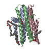
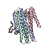
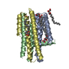
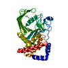
| Title | In meso in situ serial X-ray crystallography structure of diacylglycerol kinase, DgkA, at 100 K | ||||||
 Components Components | Diacylglycerol kinase | ||||||
 Keywords Keywords | TRANSFERASE | ||||||
| Function / homology |  Function and homology information Function and homology informationdiacylglycerol kinase (ATP) / lipid kinase activity / ATP-dependent diacylglycerol kinase activity / phosphatidic acid biosynthetic process / response to UV / ATP binding / membrane / metal ion binding / identical protein binding / plasma membrane Similarity search - Function | ||||||
| Biological species |  | ||||||
| Method |  X-RAY DIFFRACTION / SYNCHROTRON / MOLECULAR REPLACEMENT / Resolution: 2.8 Å X-RAY DIFFRACTION / SYNCHROTRON / MOLECULAR REPLACEMENT / Resolution: 2.8 Å | ||||||
 Authors Authors | Huang, C.-Y. / Howe, N. / Olieric, V. / Warshamanage, R. / Diederichs, K. / Wang, M. / Caffrey, M. | ||||||
| Funding support |  Ireland, 1items Ireland, 1items
| ||||||
 Citation Citation | Journal: Acta Crystallogr D Struct Biol / Year: 2016 Title: In meso in situ serial X-ray crystallography of soluble and membrane proteins at cryogenic temperatures. Authors: Huang, C.Y. / Olieric, V. / Ma, P. / Howe, N. / Vogeley, L. / Liu, X. / Warshamanage, R. / Weinert, T. / Panepucci, E. / Kobilka, B. / Diederichs, K. / Wang, M. / Caffrey, M. | ||||||
| History |
|
- Structure visualization
| Structure viewer | Molecule: MolmilJmol/JSmol |
|---|
- Downloads & links
-Download
| PDBx/mmCIF format | 5d56.cif.gz | 131.2 KB | Display | PDBx/mmCIF format |
|---|---|---|---|---|
| PDB format | pdb5d56.ent.gz | 103.4 KB | Display | PDB format |
| PDBx/mmJSON format | 5d56.json.gz | Tree view | PDBx/mmJSON format | |
| Others |  Other downloads Other downloads |
-Validation report
| Arichive directory | https://data.pdbj.org/pub/pdb/validation_reports/d5/5d56ftp://data.pdbj.org/pub/pdb/validation_reports/d5/5d56 | HTTPS FTP |
|---|
-Related structure data
| Related structure data |  5d52C  5d53C  5d54C  5d57C 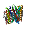 5d58C 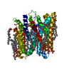 5d59C 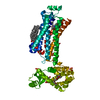 5d5aC  5d5bC 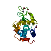 5d5cC 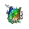 5d5dC 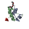 5d5eC 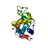 5d5fC 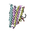 3ze3S S: Starting model for refinement C: citing same article ( |
|---|---|
| Similar structure data |


-Links
 PDBj
PDBj


- Assembly
| Deposited unit | 
| ||||||||
|---|---|---|---|---|---|---|---|---|---|
| 1 | 
| ||||||||
| 2 | 
| ||||||||
| Unit cell |
|
-Components
-Protein , 1 types, 6 molecules ABCDEF
| #1: Protein | Mass: 14204.451 Da / Num. of mol.: 6 / Mutation: A41C, C46A, I53V, I70L, M96L, V107D and C113A Source method: isolated from a genetically manipulated source Source: (gene. exp.) |
|---|
-Non-polymers , 5 types, 27 molecules 


| #2: Chemical | ChemComp-78M / (  Mass: 314.460 Da / Num. of mol.: 11 / Source method: obtained synthetically / Formula: C18H34O4 Mass: 314.460 Da / Num. of mol.: 11 / Source method: obtained synthetically / Formula: C18H34O4#3: Chemical | ChemComp-ZN / |  Mass: 65.409 Da / Num. of mol.: 1 / Source method: obtained synthetically / Formula: Zn Mass: 65.409 Da / Num. of mol.: 1 / Source method: obtained synthetically / Formula: Zn#4: Chemical | ChemComp-FLC / |  Mass: 189.100 Da / Num. of mol.: 1 / Source method: obtained synthetically / Formula: C6H5O7 Mass: 189.100 Da / Num. of mol.: 1 / Source method: obtained synthetically / Formula: C6H5O7#5: Chemical | ChemComp-ACT / |  Mass: 59.044 Da / Num. of mol.: 1 / Source method: obtained synthetically / Formula: C2H3O2 Mass: 59.044 Da / Num. of mol.: 1 / Source method: obtained synthetically / Formula: C2H3O2#6: Water | ChemComp-HOH / | Mass: 18.015 Da / Num. of mol.: 13 / Source method: isolated from a natural source / Formula: H2O |
|---|
-Experimental details
-Experiment
| Experiment | Method: X-RAY DIFFRACTION |
|---|
- Sample preparation
| Crystal | Density Matthews: 2.92 Å3/Da / Density % sol: 57.88 % |
|---|---|
| Crystal grow | Temperature: 277 K / Method: lipidic cubic phase Details: 3-6 %(v/v) MPD, 0.1 M NaCl, 60 mM Mg(CH3COO)2 and 50 mM Na3C6H5O7, pH 5.6 PH range: 5.6 |
-Data collection
| Diffraction | Mean temperature: 100 K |
|---|---|
| Diffraction source | Source: SYNCHROTRON / Site: SLS  / Beamline: X10SA / Wavelength: 1 Å / Beamline: X10SA / Wavelength: 1 Å |
| Detector | Type: DECTRIS PILATUS 6M-F / Detector: PIXEL / Date: Feb 18, 2015 |
| Radiation | Protocol: SINGLE WAVELENGTH / Monochromatic (M) / Laue (L): M / Scattering type: x-ray |
| Radiation wavelength | Wavelength: 1 Å / Relative weight: 1 |
| Reflection | Resolution: 2.8→50 Å / Num. obs: 25188 / % possible obs: 98.8 % / Redundancy: 9.3 % / Net I/σ(I): 4.42 |
| Reflection shell | Resolution: 2.8→2.87 Å / Redundancy: 8.7 % / Mean I/σ(I) obs: 1 / % possible all: 98.6 |
- Processing
| Software |
| ||||||||||||||||||||||||||||||||||||||||||||||||||||||||||||||||||||||
|---|---|---|---|---|---|---|---|---|---|---|---|---|---|---|---|---|---|---|---|---|---|---|---|---|---|---|---|---|---|---|---|---|---|---|---|---|---|---|---|---|---|---|---|---|---|---|---|---|---|---|---|---|---|---|---|---|---|---|---|---|---|---|---|---|---|---|---|---|---|---|---|
| Refinement | Method to determine structure: MOLECULAR REPLACEMENT Starting model: 3ze3 Resolution: 2.8→46.52 Å / SU ML: 0.49 / Cross valid method: THROUGHOUT / σ(F): 1.36 / Phase error: 32.94 / Stereochemistry target values: ML
| ||||||||||||||||||||||||||||||||||||||||||||||||||||||||||||||||||||||
| Solvent computation | Shrinkage radii: 0.9 Å / VDW probe radii: 1.11 Å / Solvent model: FLAT BULK SOLVENT MODEL | ||||||||||||||||||||||||||||||||||||||||||||||||||||||||||||||||||||||
| Refinement step | Cycle: LAST / Resolution: 2.8→46.52 Å
| ||||||||||||||||||||||||||||||||||||||||||||||||||||||||||||||||||||||
| Refine LS restraints |
| ||||||||||||||||||||||||||||||||||||||||||||||||||||||||||||||||||||||
| LS refinement shell |
|
