 Movie
Movie Controller

[English] 日本語
 Yorodumi
- PDB-4csi: Crystal structure of the thermostable Cellobiohydrolase Cel7A fro... -
+ Open data

- Basic information
| Entry | Database: PDB / ID: 4csi | |||||||||
|---|---|---|---|---|---|---|---|---|---|---|
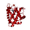
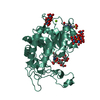
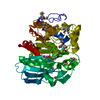
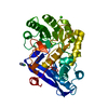
| Title | Crystal structure of the thermostable Cellobiohydrolase Cel7A from the fungus Humicola grisea var. thermoidea. | |||||||||
 Components Components | CELLULASE | |||||||||
 Keywords Keywords | HYDROLASE / GLYCOSIDE HYDROLASE | |||||||||
| Function / homology |  Function and homology information Function and homology informationcellulose 1,4-beta-cellobiosidase (non-reducing end) / cellulose 1,4-beta-cellobiosidase activity / cellulose binding / Hydrolases; Glycosylases; Glycosidases, i.e. enzymes that hydrolyse O- and S-glycosyl compounds / hydrolase activity, hydrolyzing O-glycosyl compounds / cellulose catabolic process / extracellular region Similarity search - Function | |||||||||
| Biological species |  HUMICOLA GRISEA VAR. THERMOIDEA (fungus) HUMICOLA GRISEA VAR. THERMOIDEA (fungus) | |||||||||
| Method |  X-RAY DIFFRACTION / SYNCHROTRON / MOLECULAR REPLACEMENT / Resolution: 1.8 Å X-RAY DIFFRACTION / SYNCHROTRON / MOLECULAR REPLACEMENT / Resolution: 1.8 Å | |||||||||
 Authors Authors | Haddad-Momeni, M. / Goedegebuur, F. / Hansson, H. / Karkehabadi, S. / Askarieh, G. / Mitchinson, C. / Larenas, E. / Stahlberg, J. / Sandgren, M. | |||||||||
 Citation Citation | Journal: Acta Crystallogr.,Sect.D / Year: 2014 Title: Expression, Crystal Structure and Cellulase Activity of the Thermostable Cellobiohydrolase Cel7A from the Fungus Humicola Grisea Var. Thermoidea. Authors: Haddad-Momeni, M. / Goedegebuur, F. / Hansson, H. / Karkehabadi, S. / Askarieh, G. / Mitchinson, C. / Larenas, E. / Stahlberg, J. / Sandgren, M. | |||||||||
| History |
|
- Structure visualization
| Structure viewer | Molecule: MolmilJmol/JSmol |
|---|
- Downloads & links
-Download
| PDBx/mmCIF format | 4csi.cif.gz | 191.8 KB | Display | PDBx/mmCIF format |
|---|---|---|---|---|
| PDB format | pdb4csi.ent.gz | 152.1 KB | Display | PDB format |
| PDBx/mmJSON format | 4csi.json.gz | Tree view | PDBx/mmJSON format | |
| Others |  Other downloads Other downloads |
-Validation report
| Arichive directory | https://data.pdbj.org/pub/pdb/validation_reports/cs/4csiftp://data.pdbj.org/pub/pdb/validation_reports/cs/4csi | HTTPS FTP |
|---|
-Related structure data
| Related structure data |  1celS S: Starting model for refinement |
|---|---|
| Similar structure data |


-Links
 PDBj
PDBj- Assembly
| Deposited unit | 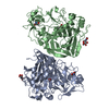
| ||||||||
|---|---|---|---|---|---|---|---|---|---|
| 1 | 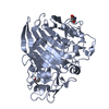
| ||||||||
| 2 | 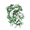
| ||||||||
| Unit cell |
|
-Components
| #1: Protein | Mass: 47113.938 Da / Num. of mol.: 2 / Fragment: CATALYTIC MODULE, RESIDUES 19-457 Source method: isolated from a genetically manipulated source Source: (gene. exp.) HUMICOLA GRISEA VAR. THERMOIDEA (fungus)Plasmid: PTREX2G / Production host: HYPOCREA JECORINA (fungus)References: UniProt: Q12621, UniProt: P15828*PLUS, cellulose 1,4-beta-cellobiosidase (reducing end) #2: Sugar |   Type: D-saccharide, beta linking / Mass: 221.208 Da / Num. of mol.: 2 Type: D-saccharide, beta linking / Mass: 221.208 Da / Num. of mol.: 2Source method: isolated from a genetically manipulated source Formula: C8H15NO6 #3: Chemical |   Mass: 106.120 Da / Num. of mol.: 2 / Source method: obtained synthetically / Formula: C4H10O3 Mass: 106.120 Da / Num. of mol.: 2 / Source method: obtained synthetically / Formula: C4H10O3#4: Water | ChemComp-HOH / |  Mass: 18.015 Da / Num. of mol.: 717 / Source method: isolated from a natural source / Formula: H2O Mass: 18.015 Da / Num. of mol.: 717 / Source method: isolated from a natural source / Formula: H2OHas protein modification | Y | Nonpolymer details | N-ACETYL-D-GLUCOSAMIN | Sequence details | THE ISOLATED GENE HAS HISTIDINE H WHEREAS Q12621 HAS TYROSINE Y AT POSITION 101 | |
|---|
-Experimental details
-Experiment
| Experiment | Method: X-RAY DIFFRACTION / Number of used crystals: 2 |
|---|
- Sample preparation
| Crystal | Density Matthews: 1.78 Å3/Da / Density % sol: 31 % / Description: NONE |
|---|---|
| Crystal grow | pH: 6 Details: PROTEIN WAS CRYSTALLIZED FROM 22 % (W/V) PEG 8000, 0.2 M AMMONIUM SULPHATE, 20 MM TRIS HCL, PH 7.0 |
-Data collection
| Diffraction | Mean temperature: 100 K |
|---|---|
| Diffraction source | Source: SYNCHROTRON / Site: ESRF  / Beamline: ID14-1 / Wavelength: 0.934 / Beamline: ID14-1 / Wavelength: 0.934 |
| Detector | Type: ADSC QUANTUM 210 / Detector: CCD / Date: Dec 1, 2004 / Details: MIRRORS |
| Radiation | Protocol: SINGLE WAVELENGTH / Monochromatic (M) / Laue (L): M / Scattering type: x-ray |
| Radiation wavelength | Wavelength: 0.934 Å / Relative weight: 1 |
| Reflection | Resolution: 1.8→34.7 Å / Num. obs: 65221 / % possible obs: 99.8 % / Observed criterion σ(I): 2 / Redundancy: 3.9 % / Rmerge(I) obs: 0.09 / Net I/σ(I): 7.1 |
| Reflection shell | Resolution: 1.8→1.9 Å / Redundancy: 3.8 % / Rmerge(I) obs: 0.41 / Mean I/σ(I) obs: 2.68 / % possible all: 99.8 |
- Processing
| Software |
| ||||||||||||||||||||||||||||||||||||||||||||||||||||||||||||||||||||||||||||||||||||||||||||||||||||||||||||||||||||||||||||||||||||||||||||||||||||||||||||||||||||||||||||||||||||||
|---|---|---|---|---|---|---|---|---|---|---|---|---|---|---|---|---|---|---|---|---|---|---|---|---|---|---|---|---|---|---|---|---|---|---|---|---|---|---|---|---|---|---|---|---|---|---|---|---|---|---|---|---|---|---|---|---|---|---|---|---|---|---|---|---|---|---|---|---|---|---|---|---|---|---|---|---|---|---|---|---|---|---|---|---|---|---|---|---|---|---|---|---|---|---|---|---|---|---|---|---|---|---|---|---|---|---|---|---|---|---|---|---|---|---|---|---|---|---|---|---|---|---|---|---|---|---|---|---|---|---|---|---|---|---|---|---|---|---|---|---|---|---|---|---|---|---|---|---|---|---|---|---|---|---|---|---|---|---|---|---|---|---|---|---|---|---|---|---|---|---|---|---|---|---|---|---|---|---|---|---|---|---|---|
| Refinement | Method to determine structure: MOLECULAR REPLACEMENT Starting model: PDB ENTRY 1CEL Resolution: 1.8→72.32 Å / Cor.coef. Fo:Fc: 0.96 / Cor.coef. Fo:Fc free: 0.938 / SU B: 2.878 / SU ML: 0.089 / Cross valid method: THROUGHOUT / ESU R: 0.144 / ESU R Free: 0.133 / Stereochemistry target values: MAXIMUM LIKELIHOOD Details: HYDROGENS HAVE BEEN ADDED IN THE RIDING POSITIONS. RESIDUES 193-200 ARE DISORDERED IN CHAIN B, RESIDUES 438 AND 439 ARE DISORDERED IN CHAIN A, AND NOT PRESENT IN THE FINAL MODEL
| ||||||||||||||||||||||||||||||||||||||||||||||||||||||||||||||||||||||||||||||||||||||||||||||||||||||||||||||||||||||||||||||||||||||||||||||||||||||||||||||||||||||||||||||||||||||
| Solvent computation | Ion probe radii: 0.8 Å / Shrinkage radii: 0.8 Å / VDW probe radii: 1.2 Å / Solvent model: MASK | ||||||||||||||||||||||||||||||||||||||||||||||||||||||||||||||||||||||||||||||||||||||||||||||||||||||||||||||||||||||||||||||||||||||||||||||||||||||||||||||||||||||||||||||||||||||
| Displacement parameters | Biso mean: 17.6 Å2
| ||||||||||||||||||||||||||||||||||||||||||||||||||||||||||||||||||||||||||||||||||||||||||||||||||||||||||||||||||||||||||||||||||||||||||||||||||||||||||||||||||||||||||||||||||||||
| Refinement step | Cycle: LAST / Resolution: 1.8→72.32 Å
| ||||||||||||||||||||||||||||||||||||||||||||||||||||||||||||||||||||||||||||||||||||||||||||||||||||||||||||||||||||||||||||||||||||||||||||||||||||||||||||||||||||||||||||||||||||||
| Refine LS restraints |
|