 Movie
Movie Controller

[English] 日本語
 Yorodumi
- PDB-1cel: THE THREE-DIMENSIONAL CRYSTAL STRUCTURE OF THE CATALYTIC CORE OF ... -
+ Open data

- Basic information
| Entry | Database: PDB / ID: 1cel | |||||||||
|---|---|---|---|---|---|---|---|---|---|---|
| Title | THE THREE-DIMENSIONAL CRYSTAL STRUCTURE OF THE CATALYTIC CORE OF CELLOBIOHYDROLASE I FROM TRICHODERMA REESEI | |||||||||
 Components Components | 1,4-BETA-D-GLUCAN CELLOBIOHYDROLASE I | |||||||||
 Keywords Keywords | HYDROLASE(O-GLYCOSYL) | |||||||||
| Function / homology |  Function and homology information Function and homology informationcellulose 1,4-beta-cellobiosidase (non-reducing end) / cellulose 1,4-beta-cellobiosidase activity / cellulose binding / cellulose catabolic process / extracellular region Similarity search - Function | |||||||||
| Biological species |  Trichoderma reesei (fungus) Trichoderma reesei (fungus) | |||||||||
| Method |  X-RAY DIFFRACTION / Resolution: 1.8 Å X-RAY DIFFRACTION / Resolution: 1.8 Å | |||||||||
 Authors Authors | Divne, C. / Jones, T.A. | |||||||||
 Citation Citation | Journal: Science / Year: 1994 Title: The three-dimensional crystal structure of the catalytic core of cellobiohydrolase I from Trichoderma reesei. Authors: Divne, C. / Stahlberg, J. / Reinikainen, T. / Ruohonen, L. / Pettersson, G. / Knowles, J.K. / Teeri, T.T. / Jones, T.A. #1: Journal: J.Mol.Biol. / Year: 1993Title: Crystallization and Preliminary X-Ray Studies on the Core Proteins of Cellobiohydrolase I and Endoglucanase I from Trichoderma Reesei Authors: Divne, C. / Sinning, I. / Stahlberg, J. / Pettersson, G. / Bailey, M. / Siika-Aho, M. / Margolles-Clark, E. / Teeri, T. / Jones, T.A. | |||||||||
| History |
| |||||||||
| Remark 700 | SHEET EACH CHAIN CONTAINS TWO LARGE ANTI-PARALLEL BETA SHEETS THAT STACK FACE-TO-FACE TO FORM A ...SHEET EACH CHAIN CONTAINS TWO LARGE ANTI-PARALLEL BETA SHEETS THAT STACK FACE-TO-FACE TO FORM A BETA SANDWICH. EACH SHEET IS BIFURCATED. IN ORDER TO REPRESENT THIS FEATURE IN THE SHEET RECORDS BELOW, TWO SHEETS ARE PRESENTED FOR EACH SHEET WHICH HAVE ONE OR MORE IDENTICAL STRANDS. SHEET 1 OF THE SANDWICH IS PRESENTED AS SHEETS *A1A* AND *A1B* FOR CHAIN A AND SHEETS *B1A* AND *B1B* FOR CHAIN B. SHEET 2 OF THE SANDWICH IS PRESENTED AS SHEETS *A2A* AND *A2B* FOR CHAIN A AND SHEETS *B2A* AND *B2B* FOR CHAIN B. THE FOLLOWING B-STRANDS ARE DISJOINT IN THE TWO SHEETS OF THE B-SANDWICH: STRAND 2 OF SHEET 1 IS DISJOINT CONSISTING OF TWO STRANDS (RESIDUES 24 - 26 AND RESIDUES 84 - 87) THAT RUN IN OPPOSITE DIRECTIONS; STRAND 3 OF SHEET 1 IS DISJOINT CONSISTING OF TWO STRANDS (RESIDUES 17 - 20 AND RESIDUES 90 - 95) THAT RUN IN OPPOSITE DIRECTIONS; STRAND 8 OF SHEET 1 IS DISJOINT CONSISTING OF TWO STRANDS (RESIDUES 309 - 313 AND RESIDUES 325 - 327) THAT RUN IN THE SAME DIRECTION; STRAND 2 OF SHEET 2 IS DISJOINT CONSISTING OF TWO STRANDS (RESIDUES 107 - 111 AND RESIDUES 119 - 121) THAT RUN IN THE SAME DIRECTION. |
- Structure visualization
| Structure viewer | Molecule: MolmilJmol/JSmol |
|---|
- Downloads & links
-Download
| PDBx/mmCIF format | 1cel.cif.gz | 185.1 KB | Display | PDBx/mmCIF format |
|---|---|---|---|---|
| PDB format | pdb1cel.ent.gz | 144.5 KB | Display | PDB format |
| PDBx/mmJSON format | 1cel.json.gz | Tree view | PDBx/mmJSON format | |
| Others |  Other downloads Other downloads |
-Validation report
| Arichive directory | https://data.pdbj.org/pub/pdb/validation_reports/ce/1celftp://data.pdbj.org/pub/pdb/validation_reports/ce/1cel | HTTPS FTP |
|---|
-Related structure data
| Similar structure data |
|---|


-Links
 PDBj
PDBj


- Assembly
| Deposited unit | 
| ||||||||
|---|---|---|---|---|---|---|---|---|---|
| 1 | 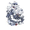
| ||||||||
| 2 | 
| ||||||||
| Unit cell |
| ||||||||
| Atom site foot note | 1: CIS PROLINE - PRO A 382 / 2: CIS PROLINE - PRO B 382 | ||||||||
| Noncrystallographic symmetry (NCS) | NCS oper: (Code: given / Matrix: (1),| Details | THE TRANSFORMATION PRESENTED ON *MTRIX* RECORDS BELOW WILL YIELD APPROXIMATE COORDINATES FOR CHAIN *B* WHEN APPLIED TO CHAIN *A*. | |
-Components
-Protein , 1 types, 2 molecules AB
| #1: Protein | Mass: 46010.703 Da / Num. of mol.: 2 Source method: isolated from a genetically manipulated source Source: (gene. exp.) Trichoderma reesei (fungus)References: UniProt: P00725, UniProt: P62694*PLUS, cellulose 1,4-beta-cellobiosidase (non-reducing end) |
|---|
-Sugars , 3 types, 4 molecules 


| #2: Sugar |  Type: D-saccharide, beta linking / Mass: 221.208 Da / Num. of mol.: 2 Type: D-saccharide, beta linking / Mass: 221.208 Da / Num. of mol.: 2Source method: isolated from a genetically manipulated source Formula: C8H15NO6 #3: Sugar | ChemComp-BGC / |  Type: D-saccharide, beta linking / Mass: 180.156 Da / Num. of mol.: 1 Type: D-saccharide, beta linking / Mass: 180.156 Da / Num. of mol.: 1Source method: isolated from a genetically manipulated source Formula: C6H12O6 #6: Sugar | ChemComp-GLC / |  Type: D-saccharide, alpha linking / Mass: 180.156 Da / Num. of mol.: 1 Type: D-saccharide, alpha linking / Mass: 180.156 Da / Num. of mol.: 1Source method: isolated from a genetically manipulated source Formula: C6H12O6 |
|---|
-Non-polymers , 3 types, 532 molecules 

| #4: Chemical | ChemComp-CA /  Mass: 40.078 Da / Num. of mol.: 1 / Source method: obtained synthetically / Formula: Ca Mass: 40.078 Da / Num. of mol.: 1 / Source method: obtained synthetically / Formula: Ca | ||
|---|---|---|---|
| #5: Chemical |  Mass: 250.100 Da / Num. of mol.: 2 / Source method: obtained synthetically / Formula: C7H7IS Mass: 250.100 Da / Num. of mol.: 2 / Source method: obtained synthetically / Formula: C7H7IS#7: Water | ChemComp-HOH / | Mass: 18.015 Da / Num. of mol.: 529 / Source method: isolated from a natural source / Formula: H2O |
-Details
| Has protein modification | Y |
|---|---|
| Nonpolymer details | A LIGAND O-IODO-BENZYL-1-THIO-B-D-GLUCOSE IS BOUND IN THE ACTIVE SITE OF EACH MOLECULE AND HAS THE ...A LIGAND O-IODO-BENZYL-1-THIO-B-D-GLUCOSE IS BOUND IN THE ACTIVE SITE OF EACH MOLECULE AND HAS THE RESIDUE NAMES IBZ AND GLC (I.E., IBZ FOR THE IODO-BENZYL GROUP AND GLC FOR THE B-D-GLUCOSE UNIT). THERE IS A CALCIUM (+II) ION COORDINATE |
| Sequence details | SEQUENCE ADVISORY NOTICE DIFFERENCE BETWEEN SWISS-PROT AND PDB SEQUENCE. SWISS-PROT ENTRY NAME: ...SEQUENCE ADVISORY NOTICE DIFFERENCE |
-Experimental details
-Experiment
| Experiment | Method: X-RAY DIFFRACTION |
|---|
- Sample preparation
| Crystal | Density Matthews: 2.2 Å3/Da / Density % sol: 44.05 % | ||||||||||||||||||||||||||||||||||||||||||
|---|---|---|---|---|---|---|---|---|---|---|---|---|---|---|---|---|---|---|---|---|---|---|---|---|---|---|---|---|---|---|---|---|---|---|---|---|---|---|---|---|---|---|---|
| Crystal grow | *PLUS pH: 3.5 / Method: vapor diffusion, hanging drop | ||||||||||||||||||||||||||||||||||||||||||
| Components of the solutions | *PLUS
|
-Data collection
| Radiation | Scattering type: x-ray |
|---|---|
| Radiation wavelength | Relative weight: 1 |
| Reflection | *PLUS Highest resolution: 1.81 Å / Num. obs: 65174 / % possible obs: 95.5 % / Num. measured all: 99089 / Rmerge(I) obs: 0.043 |
| Reflection shell | *PLUS Highest resolution: 1.81 Å / Lowest resolution: 1.87 Å / % possible obs: 52 % |
- Processing
| Software |
| ||||||||||||||||||||||||||||||||||||||||||||||||||||||||||||
|---|---|---|---|---|---|---|---|---|---|---|---|---|---|---|---|---|---|---|---|---|---|---|---|---|---|---|---|---|---|---|---|---|---|---|---|---|---|---|---|---|---|---|---|---|---|---|---|---|---|---|---|---|---|---|---|---|---|---|---|---|---|
| Refinement | Resolution: 1.8→7.5 Å / σ(F): 2 /
| ||||||||||||||||||||||||||||||||||||||||||||||||||||||||||||
| Refinement step | Cycle: LAST / Resolution: 1.8→7.5 Å
| ||||||||||||||||||||||||||||||||||||||||||||||||||||||||||||
| Refine LS restraints |
| ||||||||||||||||||||||||||||||||||||||||||||||||||||||||||||
| Software | *PLUS Name: X-PLOR / Classification: refinement | ||||||||||||||||||||||||||||||||||||||||||||||||||||||||||||
| Refinement | *PLUS Rfactor obs: 0.181 | ||||||||||||||||||||||||||||||||||||||||||||||||||||||||||||
| Solvent computation | *PLUS | ||||||||||||||||||||||||||||||||||||||||||||||||||||||||||||
| Displacement parameters | *PLUS | ||||||||||||||||||||||||||||||||||||||||||||||||||||||||||||
| Refine LS restraints | *PLUS
|
