 Movie
Movie Controller

[English] 日本語
 Yorodumi
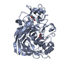
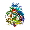
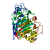
- PDB-5oa5: CELLOBIOHYDROLASE I (CEL7A) FROM HYPOCREA JECORINA WITH IMPROVED ... -
+ Open data

- Basic information
| Entry | Database: PDB / ID: 5oa5 | |||||||||
|---|---|---|---|---|---|---|---|---|---|---|
| Title | CELLOBIOHYDROLASE I (CEL7A) FROM HYPOCREA JECORINA WITH IMPROVED THERMAL STABILITY | |||||||||
 Components Components | Exoglucanase 1 | |||||||||
 Keywords Keywords | HYDROLASE / CELLULASE / PROTEIN ENGINEERING | |||||||||
| Function / homology |  Function and homology information Function and homology informationcellulose 1,4-beta-cellobiosidase (non-reducing end) / cellulose 1,4-beta-cellobiosidase activity / cellulose binding / cellulose catabolic process / extracellular region Similarity search - Function | |||||||||
| Biological species |  Hypocrea jecorina (fungus) Hypocrea jecorina (fungus) | |||||||||
| Method |  X-RAY DIFFRACTION / SYNCHROTRON / MOLECULAR REPLACEMENT / Resolution: 2.1 Å X-RAY DIFFRACTION / SYNCHROTRON / MOLECULAR REPLACEMENT / Resolution: 2.1 Å | |||||||||
 Authors Authors | Goedegebuur, F. / Hansson, H. / Karkehabadi, S. / Mikkelsen, N. / Stahlberg, J. / Sandgren, M. | |||||||||
| Funding support |  United States, 2items United States, 2items
| |||||||||
 Citation Citation | Journal: J. Biol. Chem. / Year: 2017 Title: Improving the thermal stability of cellobiohydrolase Cel7A from Hypocrea jecorina by directed evolution. Authors: Goedegebuur, F. / Dankmeyer, L. / Gualfetti, P. / Karkehabadi, S. / Hansson, H. / Jana, S. / Huynh, V. / Kelemen, B.R. / Kruithof, P. / Larenas, E.A. / Teunissen, P.J.M. / Stahlberg, J. / ...Authors: Goedegebuur, F. / Dankmeyer, L. / Gualfetti, P. / Karkehabadi, S. / Hansson, H. / Jana, S. / Huynh, V. / Kelemen, B.R. / Kruithof, P. / Larenas, E.A. / Teunissen, P.J.M. / Stahlberg, J. / Payne, C.M. / Mitchinson, C. / Sandgren, M. | |||||||||
| History |
|
- Structure visualization
| Structure viewer | Molecule: MolmilJmol/JSmol |
|---|
- Downloads & links
-Download
| PDBx/mmCIF format | 5oa5.cif.gz | 187 KB | Display | PDBx/mmCIF format |
|---|---|---|---|---|
| PDB format | pdb5oa5.ent.gz | 146.2 KB | Display | PDB format |
| PDBx/mmJSON format | 5oa5.json.gz | Tree view | PDBx/mmJSON format | |
| Others |  Other downloads Other downloads |
-Validation report
| Arichive directory | https://data.pdbj.org/pub/pdb/validation_reports/oa/5oa5ftp://data.pdbj.org/pub/pdb/validation_reports/oa/5oa5 | HTTPS FTP |
|---|
-Related structure data
| Related structure data |  2v3iS S: Starting model for refinement |
|---|---|
| Similar structure data |


-Links
 PDBj
PDBj- Assembly
| Deposited unit | 
| ||||||||
|---|---|---|---|---|---|---|---|---|---|
| 1 | 
| ||||||||
| 2 | 
| ||||||||
| Unit cell |
|
-Components
| #1: Protein | Mass: 46262.238 Da / Num. of mol.: 2 / Fragment: CATALYTIC DOMAIN, RESIDUES 18-451 Mutation: S8P, T41I, N49S, A68T, N89D, S92T, S113N, S196T, P227L, D249K, T255P, S278P, E295K, T296P, T332Y, V304D, S411F Source method: isolated from a genetically manipulated source Source: (gene. exp.) Hypocrea jecorina (fungus) / Gene: cbh1 / Plasmid: pRAXDES / Production host: References: UniProt: P62694, cellulose 1,4-beta-cellobiosidase (non-reducing end) #2: Sugar | ChemComp-NAG /   Type: D-saccharide, beta linking / Mass: 221.208 Da / Num. of mol.: 6 Type: D-saccharide, beta linking / Mass: 221.208 Da / Num. of mol.: 6Source method: isolated from a genetically manipulated source Formula: C8H15NO6 #3: Chemical | ChemComp-GOL / |   Mass: 92.094 Da / Num. of mol.: 1 / Source method: obtained synthetically / Formula: C3H8O3 Mass: 92.094 Da / Num. of mol.: 1 / Source method: obtained synthetically / Formula: C3H8O3#4: Water | ChemComp-HOH / |  Mass: 18.015 Da / Num. of mol.: 546 / Source method: isolated from a natural source / Formula: H2O Mass: 18.015 Da / Num. of mol.: 546 / Source method: isolated from a natural source / Formula: H2OHas protein modification | Y | Sequence details | ENGINEERED | |
|---|
-Experimental details
-Experiment
| Experiment | Method: X-RAY DIFFRACTION / Number of used crystals: 1 |
|---|
- Sample preparation
| Crystal | Density Matthews: 2.48 Å3/Da / Density % sol: 50.45 % |
|---|---|
| Crystal grow | Temperature: 293 K / Method: vapor diffusion, sitting drop / pH: 6 Details: 25.5% POLYETHYLENE GLYCOL (PEG) 4000, 0.17 M AMSO4 AND 15% GLYCEROL |
-Data collection
| Diffraction | Mean temperature: 100 K |
|---|---|
| Diffraction source | Source: SYNCHROTRON / Site: ESRF  / Beamline: ID23-2 / Wavelength: 0.8726 Å / Beamline: ID23-2 / Wavelength: 0.8726 Å |
| Detector | Type: MARRESEARCH / Detector: CCD / Date: Oct 5, 2012 / Details: PT COATED MIRRORS |
| Radiation | Monochromator: HORIZONTALLY DIFFRACTING SI (111) MONOCHROMATOR Protocol: SINGLE WAVELENGTH / Monochromatic (M) / Laue (L): M / Scattering type: x-ray |
| Radiation wavelength | Wavelength: 0.8726 Å / Relative weight: 1 |
| Reflection | Resolution: 1.99→41.16 Å / Num. obs: 56990 / % possible obs: 99.6 % / Observed criterion σ(I): 2 / Redundancy: 3.71 % / Rmerge(I) obs: 0.13 / Net I/σ(I): 4.9 |
| Reflection shell | Resolution: 2.1→2.21 Å / Redundancy: 3.73 % / Rmerge(I) obs: 0.41 / Mean I/σ(I) obs: 1.86 / % possible all: 99.8 |
- Processing
| Software |
| ||||||||||||||||||||||||||||||||||||||||||||||||||||||||||||||||||||||||||||||||||||||||||||||||||||||||||||||||||||||||||||||||||||||||||||||||||||||||||||||||||||||||||||||||||||||
|---|---|---|---|---|---|---|---|---|---|---|---|---|---|---|---|---|---|---|---|---|---|---|---|---|---|---|---|---|---|---|---|---|---|---|---|---|---|---|---|---|---|---|---|---|---|---|---|---|---|---|---|---|---|---|---|---|---|---|---|---|---|---|---|---|---|---|---|---|---|---|---|---|---|---|---|---|---|---|---|---|---|---|---|---|---|---|---|---|---|---|---|---|---|---|---|---|---|---|---|---|---|---|---|---|---|---|---|---|---|---|---|---|---|---|---|---|---|---|---|---|---|---|---|---|---|---|---|---|---|---|---|---|---|---|---|---|---|---|---|---|---|---|---|---|---|---|---|---|---|---|---|---|---|---|---|---|---|---|---|---|---|---|---|---|---|---|---|---|---|---|---|---|---|---|---|---|---|---|---|---|---|---|---|
| Refinement | Method to determine structure: MOLECULAR REPLACEMENT Starting model: PDB ENTRY 2V3I Resolution: 2.1→41.16 Å / Cor.coef. Fo:Fc: 0.951 / Cor.coef. Fo:Fc free: 0.926 / SU B: 4.952 / SU ML: 0.13 / Cross valid method: THROUGHOUT / ESU R: 0.226 / ESU R Free: 0.19 / Details: HYDROGENS HAVE BEEN USED IF PRESENT IN THE INPUT
| ||||||||||||||||||||||||||||||||||||||||||||||||||||||||||||||||||||||||||||||||||||||||||||||||||||||||||||||||||||||||||||||||||||||||||||||||||||||||||||||||||||||||||||||||||||||
| Solvent computation | Ion probe radii: 0.8 Å / Shrinkage radii: 0.8 Å / VDW probe radii: 1.2 Å | ||||||||||||||||||||||||||||||||||||||||||||||||||||||||||||||||||||||||||||||||||||||||||||||||||||||||||||||||||||||||||||||||||||||||||||||||||||||||||||||||||||||||||||||||||||||
| Displacement parameters | Biso mean: 17.9 Å2
| ||||||||||||||||||||||||||||||||||||||||||||||||||||||||||||||||||||||||||||||||||||||||||||||||||||||||||||||||||||||||||||||||||||||||||||||||||||||||||||||||||||||||||||||||||||||
| Refinement step | Cycle: LAST / Resolution: 2.1→41.16 Å
| ||||||||||||||||||||||||||||||||||||||||||||||||||||||||||||||||||||||||||||||||||||||||||||||||||||||||||||||||||||||||||||||||||||||||||||||||||||||||||||||||||||||||||||||||||||||
| Refine LS restraints |
|