 Movie
Movie Controller


+ Open data

- Basic information
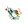
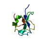
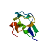
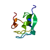
| Entry | Database: PDB / ID: 1ucs | ||||||
|---|---|---|---|---|---|---|---|
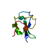
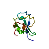
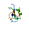
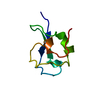
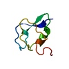
| Title | Type III Antifreeze Protein RD1 from an Antarctic Eel Pout | ||||||
 Components Components | Antifreeze peptide RD1 | ||||||
 Keywords Keywords | ANTIFREEZE PROTEIN / Small Beta Barrel / Pretzel Fold | ||||||
| Function / homology |  Function and homology information Function and homology information | ||||||
| Biological species |  Lycodichthys dearborni (Antarctic eel pout) Lycodichthys dearborni (Antarctic eel pout) | ||||||
| Method |  X-RAY DIFFRACTION / SYNCHROTRON / MOLECULAR REPLACEMENT / Resolution: 0.62 Å X-RAY DIFFRACTION / SYNCHROTRON / MOLECULAR REPLACEMENT / Resolution: 0.62 Å | ||||||
 Authors Authors | Ko, T.-P. / Robinson, H. / Gao, Y.-G. / Cheng, C.-H.C. / DeVries, A.L. / Wang, A.H.-J. | ||||||
 Citation Citation | Journal: Biophys.J. / Year: 2003 Title: The refined crystal structure of an eel pout type III antifreeze protein RD1 at 0.62-A resolution reveals structural microheterogeneity of protein and solvation. Authors: Ko, T.P. / Robinson, H. / Gao, Y.G. / Cheng, C.H. / DeVries, A.L. / Wang, A.H. | ||||||
| History |
|
- Structure visualization
| Structure viewer | Molecule: MolmilJmol/JSmol |
|---|
- Downloads & links
-Download
| PDBx/mmCIF format | 1ucs.cif.gz | 39.3 KB | Display | PDBx/mmCIF format |
|---|---|---|---|---|
| PDB format | pdb1ucs.ent.gz | 28.1 KB | Display | PDB format |
| PDBx/mmJSON format | 1ucs.json.gz | Tree view | PDBx/mmJSON format | |
| Others |  Other downloads Other downloads |
-Validation report
| Arichive directory | https://data.pdbj.org/pub/pdb/validation_reports/uc/1ucsftp://data.pdbj.org/pub/pdb/validation_reports/uc/1ucs | HTTPS FTP |
|---|
-Related structure data
| Related structure data | |
|---|---|
| Similar structure data |


-Links
 PDBj
PDBj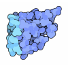
- Assembly
| Deposited unit | 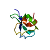
| ||||||||
|---|---|---|---|---|---|---|---|---|---|
| 1 |
| ||||||||
| Unit cell |
|
-Components
| #1: Protein | Mass: 6911.279 Da / Num. of mol.: 1 / Source method: isolated from a natural source Source: (natural) Lycodichthys dearborni (Antarctic eel pout)References: UniProt: P35751 |
|---|---|
| #2: Water | ChemComp-HOH /  Mass: 18.015 Da / Num. of mol.: 240 / Source method: isolated from a natural source / Formula: H2O Mass: 18.015 Da / Num. of mol.: 240 / Source method: isolated from a natural source / Formula: H2O |
-Experimental details
-Experiment
| Experiment | Method: X-RAY DIFFRACTION / Number of used crystals: 1 |
|---|
- Sample preparation
| Crystal | Density Matthews: 1.4 Å3/Da / Density % sol: 11.58 % | ||||||||||||||||||||||||||||||
|---|---|---|---|---|---|---|---|---|---|---|---|---|---|---|---|---|---|---|---|---|---|---|---|---|---|---|---|---|---|---|---|
| Crystal grow | Temperature: 298 K / Method: vapor diffusion, hanging drop / pH: 7.5 Details: Ammonium Sulfate, Tris-HCl, pH 7.5, VAPOR DIFFUSION, HANGING DROP, temperature 298.0K | ||||||||||||||||||||||||||||||
| Crystal grow | *PLUS | ||||||||||||||||||||||||||||||
| Components of the solutions | *PLUS
|
-Data collection
| Diffraction | Mean temperature: 110 K |
|---|---|
| Diffraction source | Source: SYNCHROTRON / Site: APS  / Beamline: 19-ID / Wavelength: 0.6668 Å / Beamline: 19-ID / Wavelength: 0.6668 Å |
| Detector | Type: CUSTOM-MADE / Detector: CCD / Date: Jan 23, 2000 |
| Radiation | Monochromator: Si 111 CHANNEL / Protocol: SINGLE WAVELENGTH / Monochromatic (M) / Laue (L): M / Scattering type: x-ray |
| Radiation wavelength | Wavelength: 0.6668 Å / Relative weight: 1 |
| Reflection | Resolution: 0.62→50 Å / Num. all: 127366 / Num. obs: 118502 / % possible obs: 94 % / Observed criterion σ(F): 0 / Observed criterion σ(I): 1 / Redundancy: 3.3 % / Rmerge(I) obs: 0.072 / Net I/σ(I): 21.5 |
| Reflection shell | Resolution: 0.62→0.64 Å / Redundancy: 1.9 % / Rmerge(I) obs: 0.653 / Mean I/σ(I) obs: 1.2 / Num. unique all: 10198 / % possible all: 91.8 |
| Reflection | *PLUS Lowest resolution: 1.2 Å / Num. obs: 108526 / % possible obs: 97 % / Num. measured all: 256420 / Rmerge(I) obs: 0.073 |
| Reflection shell | *PLUS % possible obs: 91.8 % / Num. unique obs: 10198 / Num. measured obs: 18801 |
- Processing
| Software |
| |||||||||||||||||||||||||
|---|---|---|---|---|---|---|---|---|---|---|---|---|---|---|---|---|---|---|---|---|---|---|---|---|---|---|
| Refinement | Method to determine structure: MOLECULAR REPLACEMENT Starting model: A polypeptide model derived from NMR experiments Resolution: 0.62→22.32 Å / Isotropic thermal model: anisotropic / Cross valid method: THROUGHOUT / σ(F): 0 / σ(I): 0 / Stereochemistry target values: Engh & Huber Details: anisotropic model was used in refinement and then the converted to isotropic B values
| |||||||||||||||||||||||||
| Displacement parameters | Biso mean: 8.91 Å2 | |||||||||||||||||||||||||
| Refinement step | Cycle: LAST / Resolution: 0.62→22.32 Å
| |||||||||||||||||||||||||
| Refine LS restraints | Type: s_bond_d / Dev ideal: 0.012 | |||||||||||||||||||||||||
| LS refinement shell | Resolution: 0.62→0.65 Å / Rfactor Rfree error: 0.015
| |||||||||||||||||||||||||
| Software | *PLUS Name: SHELXL / Version: 97 / Classification: refinement | |||||||||||||||||||||||||
| Refinement | *PLUS Lowest resolution: 50 Å / % reflection Rfree: 5 % | |||||||||||||||||||||||||
| Solvent computation | *PLUS | |||||||||||||||||||||||||
| Displacement parameters | *PLUS | |||||||||||||||||||||||||
| Refine LS restraints | *PLUS
|
