 Movie
Movie Controller

[English] 日本語
 Yorodumi
- PDB-1hg7: High resolution structure of HPLC-12 type III antifreeze protein ... -
+ Open data

- Basic information
| Entry | Database: PDB / ID: 1hg7 | ||||||
|---|---|---|---|---|---|---|---|
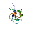
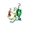
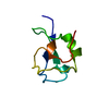
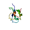
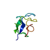
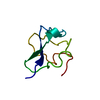
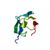
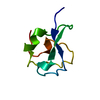
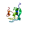
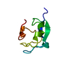
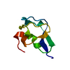
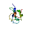
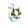
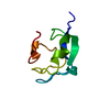
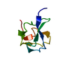
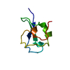
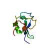
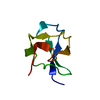
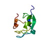
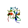
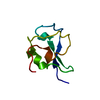
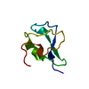
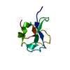
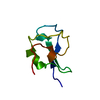
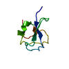
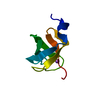
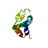
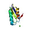
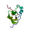
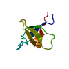
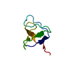
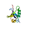
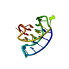
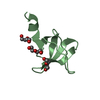
| Title | High resolution structure of HPLC-12 type III antifreeze protein from Ocean Pout Macrozoarces americanus | ||||||
 Components Components | HPLC-12 TYPE III ANTIFREEZE PROTEIN | ||||||
 Keywords Keywords | ANTIFREEZE PROTEIN / OCEAN POUT / MACROZOARCES AMERICANUS / ICE-BINDING PROTEIN | ||||||
| Function / homology |  Function and homology information Function and homology information | ||||||
| Biological species |  MACROZOARCES AMERICANUS (ocean pout) MACROZOARCES AMERICANUS (ocean pout) | ||||||
| Method |  X-RAY DIFFRACTION / SYNCHROTRON / MOLECULAR REPLACEMENT / Resolution: 1.15 Å X-RAY DIFFRACTION / SYNCHROTRON / MOLECULAR REPLACEMENT / Resolution: 1.15 Å | ||||||
 Authors Authors | Antson, A.A. / Smith, D.J. / Roper, D.I. / Lewis, S. / Caves, L.S.D. / Verma, C.S. / Buckley, S.L. / Lillford, P.J. / Hubbard, R.E. | ||||||
 Citation Citation | Journal: J.Mol.Biol. / Year: 2001 Title: Understanding the Mechanism of Ice Binding by Type III Antifreeze Protein Authors: Antson, A.A. / Smith, D.J. / Roper, D.I. / Lewis, S. / Caves, L.S.D. / Verma, C.S. / Buckley, S.L. / Lillford, P.J. / Hubbard, R.E. | ||||||
| History |
|
- Structure visualization
| Structure viewer | Molecule: MolmilJmol/JSmol |
|---|
- Downloads & links
-Download
| PDBx/mmCIF format | 1hg7.cif.gz | 46.4 KB | Display | PDBx/mmCIF format |
|---|---|---|---|---|
| PDB format | pdb1hg7.ent.gz | 32.4 KB | Display | PDB format |
| PDBx/mmJSON format | 1hg7.json.gz | Tree view | PDBx/mmJSON format | |
| Others |  Other downloads Other downloads |
-Validation report
| Arichive directory | https://data.pdbj.org/pub/pdb/validation_reports/hg/1hg7ftp://data.pdbj.org/pub/pdb/validation_reports/hg/1hg7 | HTTPS FTP |
|---|
-Related structure data
| Related structure data | 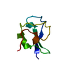 1gziS S: Starting model for refinement |
|---|---|
| Similar structure data |


-Links
 PDBj
PDBj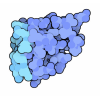
- Assembly
| Deposited unit | 
| ||||||||
|---|---|---|---|---|---|---|---|---|---|
| 1 |
| ||||||||
| Unit cell |
|
-Components
| #1: Protein | Mass: 7039.351 Da / Num. of mol.: 1 / Mutation: YES Source method: isolated from a genetically manipulated source Source: (gene. exp.) MACROZOARCES AMERICANUS (ocean pout) / Tissue: BLOOD SERUM / Gene: RECOMBINANT TYPE III AFP HPURCE 10 FRACTION 12 / Variant: HPLC-12 COMPONENT / Plasmid: PET22BGene (production host): RECOMBINANT TYPE III AFP HPURCE 10 FRACTION 12 Production host:  |
|---|---|
| #2: Chemical | ChemComp-SO4 /   Mass: 96.063 Da / Num. of mol.: 1 / Source method: obtained synthetically / Formula: SO4 Mass: 96.063 Da / Num. of mol.: 1 / Source method: obtained synthetically / Formula: SO4 |
| #3: Water | ChemComp-HOH /  Mass: 18.015 Da / Num. of mol.: 154 / Source method: isolated from a natural source / Formula: H2O Mass: 18.015 Da / Num. of mol.: 154 / Source method: isolated from a natural source / Formula: H2O |
| Compound details | CHAIN A ENGINEERED MUTATION PRO64ALA, PRO65ALA, ALA66 DELETION MUTATNT ANTIFREEZE PROTEINS LOWER ...CHAIN A ENGINEERED |
-Experimental details
-Experiment
| Experiment | Method: X-RAY DIFFRACTION / Number of used crystals: 1 |
|---|
- Sample preparation
| Crystal | Density Matthews: 2.06 Å3/Da / Density % sol: 39.8 % | ||||||||||||||||||||
|---|---|---|---|---|---|---|---|---|---|---|---|---|---|---|---|---|---|---|---|---|---|
| Crystal grow | pH: 4.6 / Details: 50% AMMONIUM SULFATE, 0.1M SODIUM ACETATE, PH 4.6 | ||||||||||||||||||||
| Crystal | *PLUS Density % sol: 40.2 % | ||||||||||||||||||||
| Crystal grow | *PLUS Temperature: 20 ℃ / Method: vapor diffusion, hanging drop / PH range low: 5 / PH range high: 4.6 | ||||||||||||||||||||
| Components of the solutions | *PLUS
|
-Data collection
| Diffraction | Mean temperature: 110 K |
|---|---|
| Diffraction source | Source: SYNCHROTRON / Site: EMBL/DESY, HAMBURG  / Beamline: BW7A / Wavelength: 0.91 / Beamline: BW7A / Wavelength: 0.91 |
| Detector | Type: MARRESEARCH / Detector: IMAGE PLATE / Date: Oct 15, 1996 |
| Radiation | Protocol: SINGLE WAVELENGTH / Monochromatic (M) / Laue (L): M / Scattering type: x-ray |
| Radiation wavelength | Wavelength: 0.91 Å / Relative weight: 1 |
| Reflection | Resolution: 1.15→45 Å / Num. obs: 20883 / % possible obs: 99.9 % / Observed criterion σ(I): 0 / Redundancy: 6.7 % / Rmerge(I) obs: 0.108 / Net I/σ(I): 5.2 |
| Reflection shell | Resolution: 1.15→1.17 Å / Redundancy: 5 % / Rmerge(I) obs: 0.493 / Mean I/σ(I) obs: 3.7 / % possible all: 100 |
| Reflection | *PLUS Lowest resolution: 45 Å |
| Reflection shell | *PLUS % possible obs: 100 % |
- Processing
| Software |
| |||||||||||||||||||||||||||||||||
|---|---|---|---|---|---|---|---|---|---|---|---|---|---|---|---|---|---|---|---|---|---|---|---|---|---|---|---|---|---|---|---|---|---|---|
| Refinement | Method to determine structure: MOLECULAR REPLACEMENT Starting model: PDB ENTRY 1GZI Resolution: 1.15→40 Å / σ(F): 0
| |||||||||||||||||||||||||||||||||
| Refinement step | Cycle: LAST / Resolution: 1.15→40 Å
| |||||||||||||||||||||||||||||||||
| Refine LS restraints |
| |||||||||||||||||||||||||||||||||
| Software | *PLUS Name: SHELXL / Classification: refinement | |||||||||||||||||||||||||||||||||
| Refinement | *PLUS σ(F): 4 / Num. reflection Rfree: 2066 / Rfactor all: 0.12 / Rfactor obs: 0.104 / Rfactor Rwork: 0.12 | |||||||||||||||||||||||||||||||||
| Solvent computation | *PLUS | |||||||||||||||||||||||||||||||||
| Displacement parameters | *PLUS |
