 Movie
Movie Controller


+ Open data

- Basic information
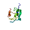
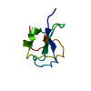
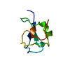
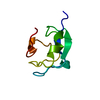
| Entry | Database: PDB / ID: 5msi | ||||||
|---|---|---|---|---|---|---|---|
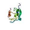
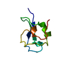
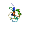
| Title | TYPE III ANTIFREEZE PROTEIN ISOFORM HPLC 12 | ||||||
 Components Components | TYPE III ANTIFREEZE PROTEIN ISOFORM HPLC 12 | ||||||
 Keywords Keywords | ANTIFREEZE / ANTIFREEZE POLYPEPTIDE / MUTANT / ICE BINDING PROTEIN / THERMAL HYSTERESIS PROTEIN / GLYCOPROTEIN | ||||||
| Function / homology |  Function and homology information Function and homology information | ||||||
| Biological species |  Macrozoarces americanus (ocean pout) Macrozoarces americanus (ocean pout) | ||||||
| Method |  X-RAY DIFFRACTION / ISOMORPHOUS / Resolution: 1.6 Å X-RAY DIFFRACTION / ISOMORPHOUS / Resolution: 1.6 Å | ||||||
 Authors Authors | Deluca, C.I. / Davies, P.L. / Ye, Q. / Jia, Z. | ||||||
 Citation Citation | Journal: J.Mol.Biol. / Year: 1998 Title: The effects of steric mutations on the structure of type III antifreeze protein and its interaction with ice. Authors: DeLuca, C.I. / Davies, P.L. / Ye, Q. / Jia, Z. | ||||||
| History |
|
- Structure visualization
| Structure viewer | Molecule: MolmilJmol/JSmol |
|---|
- Downloads & links
-Download
| PDBx/mmCIF format | 5msi.cif.gz | 24 KB | Display | PDBx/mmCIF format |
|---|---|---|---|---|
| PDB format | pdb5msi.ent.gz | 15.3 KB | Display | PDB format |
| PDBx/mmJSON format | 5msi.json.gz | Tree view | PDBx/mmJSON format | |
| Others |  Other downloads Other downloads |
-Validation report
| Arichive directory | https://data.pdbj.org/pub/pdb/validation_reports/ms/5msiftp://data.pdbj.org/pub/pdb/validation_reports/ms/5msi | HTTPS FTP |
|---|
-Related structure data
| Related structure data | 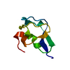 2msiC  3msiC 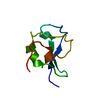 4msiC 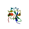 6msiC  7msiC 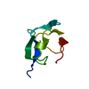 1msiS S: Starting model for refinement C: citing same article ( |
|---|---|
| Similar structure data |


-Links
 PDBj
PDBj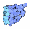
- Assembly
| Deposited unit | 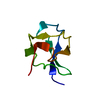
| ||||||||
|---|---|---|---|---|---|---|---|---|---|
| 1 |
| ||||||||
| Unit cell |
|
-Components
| #1: Protein | Mass: 7028.391 Da / Num. of mol.: 1 / Mutation: A16C Source method: isolated from a genetically manipulated source Source: (gene. exp.) Macrozoarces americanus (ocean pout) / Plasmid: PT7-7F-PGP1-2 / Production host:  |
|---|---|
| #2: Water | ChemComp-HOH /  Mass: 18.015 Da / Num. of mol.: 70 / Source method: isolated from a natural source / Formula: H2O Mass: 18.015 Da / Num. of mol.: 70 / Source method: isolated from a natural source / Formula: H2O |
-Experimental details
-Experiment
| Experiment | Method: X-RAY DIFFRACTION / Number of used crystals: 1 |
|---|
- Sample preparation
| Crystal | Density Matthews: 2.1 Å3/Da / Density % sol: 30 % | ||||||||||||||||||||||||||||||
|---|---|---|---|---|---|---|---|---|---|---|---|---|---|---|---|---|---|---|---|---|---|---|---|---|---|---|---|---|---|---|---|
| Crystal grow | pH: 4.5 Details: PROTEIN WAS CRYSTALLIZED IN 50-55% AMMONIUM SULFATE, 0.1 M SODIUM ACETATE PH 4-4.5 PH range: 4-4.5 | ||||||||||||||||||||||||||||||
| Crystal grow | *PLUS Temperature: 20 ℃ / Method: vapor diffusion, hanging drop / Details: Jia, Z.,(1995) Protein Sci., 4, 1236. / PH range low: 4.5 / PH range high: 4 | ||||||||||||||||||||||||||||||
| Components of the solutions | *PLUS
|
-Data collection
| Diffraction | Mean temperature: 295 K |
|---|---|
| Diffraction source | Source: ROTATING ANODE / Type: RIGAKU RUH2R / Wavelength: 1.5418 |
| Detector | Type: MARRESEARCH / Detector: IMAGE PLATE / Date: Jan 26, 1996 |
| Radiation | Monochromator: GRAPHITE(002) / Monochromatic (M) / Laue (L): M / Scattering type: x-ray |
| Radiation wavelength | Wavelength: 1.5418 Å / Relative weight: 1 |
| Reflection | Resolution: 1.6→50 Å / Num. obs: 7177 / % possible obs: 84.5 % / Observed criterion σ(I): 1 / Redundancy: 2.7 % / Rsym value: 0.066 / Net I/σ(I): 17 |
| Reflection shell | Resolution: 1.6→1.67 Å / Mean I/σ(I) obs: 2.8 / Rsym value: 0.254 / % possible all: 67.9 |
| Reflection | *PLUS Num. measured all: 19644 / Rmerge(I) obs: 0.062 |
- Processing
| Software |
| ||||||||||||||||||||
|---|---|---|---|---|---|---|---|---|---|---|---|---|---|---|---|---|---|---|---|---|---|
| Refinement | Method to determine structure: ISOMORPHOUS Starting model: 1MSI Resolution: 1.6→50 Å / Cross valid method: FREE-R REFINEMENT / σ(F): 1
| ||||||||||||||||||||
| Refinement step | Cycle: LAST / Resolution: 1.6→50 Å
| ||||||||||||||||||||
| LS refinement shell | Resolution: 1.6→1.67 Å / Total num. of bins used: 8 / % reflection obs: 93.94 % | ||||||||||||||||||||
| Xplor file |
| ||||||||||||||||||||
| Software | *PLUS Name: X-PLOR / Version: 3 / Classification: refinement | ||||||||||||||||||||
| Refinement | *PLUS Lowest resolution: 8 Å / Num. reflection obs: 7177 / Rfactor obs: 0.194 / Rfactor Rfree: 0.268 | ||||||||||||||||||||
| Solvent computation | *PLUS | ||||||||||||||||||||
| Displacement parameters | *PLUS |
