 Movie
Movie Controller

[English] 日本語
 Yorodumi
- PDB-1gzi: CRYSTAL STRUCTURE OF TYPE III ANTIFREEZE PROTEIN FROM OCEAN POUT,... -
+ Open data

- Basic information
| Entry | Database: PDB / ID: 1gzi | ||||||
|---|---|---|---|---|---|---|---|
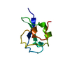
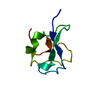
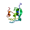
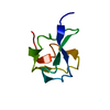
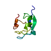
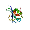
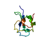
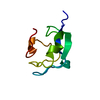
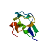
| Title | CRYSTAL STRUCTURE OF TYPE III ANTIFREEZE PROTEIN FROM OCEAN POUT, AT 1.8 ANGSTROM RESOLUTION | ||||||
 Components Components | HPLC-12 TYPE III ANTIFREEZE PROTEIN | ||||||
 Keywords Keywords | ICE-BINDING PROTEIN / ANTIFREEZE PROTEIN / OCEAN POUT / GLYCOPROTEIN / MACROZOARCES AMERICANUS / MULTIGENE FAMILY | ||||||
| Function / homology |  Function and homology information Function and homology information | ||||||
| Biological species |  Macrozoarces americanus (ocean pout) Macrozoarces americanus (ocean pout) | ||||||
| Method |  X-RAY DIFFRACTION / MIR / Resolution: 1.8 Å X-RAY DIFFRACTION / MIR / Resolution: 1.8 Å | ||||||
 Authors Authors | Antson, A.A. / Lewis, S. / Roper, D.I. / Smith, D.J. / Hubbard, R.E. | ||||||
 Citation Citation | Journal: To be Published Title: The Structure of Type III Antifreeze Protein from Ocean Pout Authors: Antson, A.A. / Lewis, S. / Roper, D.I. / Smith, D.J. / Hubbard, R.E. #1: Journal: Protein Sci. / Year: 1995Title: Crystallization and Preliminary X-Ray Crystallographic Studies on Type III Antifreeze Protein Authors: Jia, Z. / Deluca, C.I. / Davies, P.L. #2: Journal: Protein Sci. / Year: 1993Title: Use of Proline Mutants to Help Solve the NMR Solution Structure of Type III Antifreeze Protein Authors: Chao, H. / Davies, P.L. / Sykes, B.D. / Sonnichsen, F.D. | ||||||
| History |
|
- Structure visualization
| Structure viewer | Molecule: MolmilJmol/JSmol |
|---|
- Downloads & links
-Download
| PDBx/mmCIF format | 1gzi.cif.gz | 23.7 KB | Display | PDBx/mmCIF format |
|---|---|---|---|---|
| PDB format | pdb1gzi.ent.gz | 15.1 KB | Display | PDB format |
| PDBx/mmJSON format | 1gzi.json.gz | Tree view | PDBx/mmJSON format | |
| Others |  Other downloads Other downloads |
-Validation report
| Arichive directory | https://data.pdbj.org/pub/pdb/validation_reports/gz/1gziftp://data.pdbj.org/pub/pdb/validation_reports/gz/1gzi | HTTPS FTP |
|---|
-Related structure data
| Similar structure data |
|---|

-Links
 PDBj
PDBj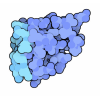
- Assembly
| Deposited unit | 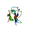
| ||||||||
|---|---|---|---|---|---|---|---|---|---|
| 1 |
| ||||||||
| Unit cell |
|
-Components
| #1: Protein | Mass: 6908.155 Da / Num. of mol.: 1 / Mutation: P64A, P65A, DEL(A66) Source method: isolated from a genetically manipulated source Source: (gene. exp.) Macrozoarces americanus (ocean pout) / Tissue: BLOOD SERUM / Cell line: BL21 / Gene: RECOMBINANT TYPE III AFP HPLC / Organ: BLOOD / Variant: HPLC-12 COMPONENT / Plasmid: PET22B / Species (production host): Escherichia coliGene (production host): RECOMBINANT TYPE III AFP HPLC FRACTION 12 Production host:  |
|---|---|
| #2: Water | ChemComp-HOH /  Mass: 18.015 Da / Num. of mol.: 54 / Source method: isolated from a natural source / Formula: H2O Mass: 18.015 Da / Num. of mol.: 54 / Source method: isolated from a natural source / Formula: H2O |
-Experimental details
-Experiment
| Experiment | Method: X-RAY DIFFRACTION / Number of used crystals: 1 |
|---|
- Sample preparation
| Crystal | Density Matthews: 2.06 Å3/Da / Density % sol: 39.8 % |
|---|---|
| Crystal grow | pH: 5.6 / Details: 50% AMMONIUM SULFATE, 0.1M SODIUM ACETATE, PH 5.6 |
-Data collection
| Diffraction | Mean temperature: 293 K |
|---|---|
| Diffraction source | Source: ROTATING ANODE / Type: RIGAKU RUH2R / Wavelength: 1.5418 |
| Detector | Type: MARRESEARCH / Detector: IMAGE PLATE / Date: Jul 18, 1996 |
| Radiation | Monochromator: GRAPHITE(002) / Monochromatic (M) / Laue (L): M / Scattering type: x-ray |
| Radiation wavelength | Wavelength: 1.5418 Å / Relative weight: 1 |
| Reflection | Resolution: 1.8→15 Å / Num. obs: 5155 / % possible obs: 99.7 % / Observed criterion σ(I): 0 / Redundancy: 3.6 % / Biso Wilson estimate: 21.2 Å2 / Rmerge(I) obs: 0.065 / Net I/σ(I): 6.3 |
| Reflection shell | Resolution: 1.8→1.9 Å / Redundancy: 3.6 % / Rmerge(I) obs: 0.208 / Mean I/σ(I) obs: 3.2 / % possible all: 100 |
- Processing
| Software |
| ||||||||||||||||||||||||||||||||||||||||||||||||||||||||||||||||||||||||||||||||||||
|---|---|---|---|---|---|---|---|---|---|---|---|---|---|---|---|---|---|---|---|---|---|---|---|---|---|---|---|---|---|---|---|---|---|---|---|---|---|---|---|---|---|---|---|---|---|---|---|---|---|---|---|---|---|---|---|---|---|---|---|---|---|---|---|---|---|---|---|---|---|---|---|---|---|---|---|---|---|---|---|---|---|---|---|---|---|
| Refinement | Method to determine structure: MIR / Resolution: 1.8→15 Å / σ(F): 0 Details: THE FOLLOWING RESIDUES WERE MODELED IN TWO CONFORMATIONS: VAL A 20 - SIDE CHAIN ATOMS. SER A 42 - TWO CONFORMATIONS FOR THE SIDE CHAIN HYDROXYL.
| ||||||||||||||||||||||||||||||||||||||||||||||||||||||||||||||||||||||||||||||||||||
| Displacement parameters | Biso mean: 25.3 Å2 | ||||||||||||||||||||||||||||||||||||||||||||||||||||||||||||||||||||||||||||||||||||
| Refinement step | Cycle: LAST / Resolution: 1.8→15 Å
| ||||||||||||||||||||||||||||||||||||||||||||||||||||||||||||||||||||||||||||||||||||
| Refine LS restraints |
|
