 Movie
Movie Controller

[English] 日本語
 Yorodumi
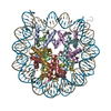
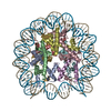
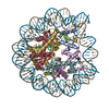
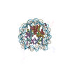
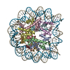
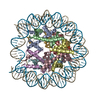
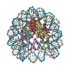
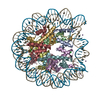
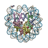
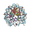
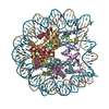
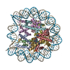
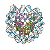
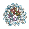
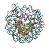
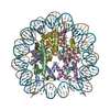
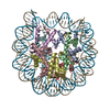
- PDB-3c1b: The effect of H3 K79 dimethylation and H4 K20 trimethylation on n... -
+ Open data

- Basic information
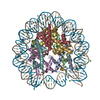
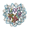
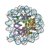
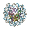
| Entry | Database: PDB / ID: 3c1b | ||||||
|---|---|---|---|---|---|---|---|
| Title | The effect of H3 K79 dimethylation and H4 K20 trimethylation on nucleosome and chromatin structure | ||||||
 Components Components |
| ||||||
 Keywords Keywords | Structural PROTEIN/DNA / Nucleosome / Dimethylated histone / trimethylated histone / methylation / nucleosomal surface / histone modification / nucleosomal array / chromatin / Acetylation / Chromosomal protein / DNA-binding / Nucleosome core / Nucleus / Phosphoprotein / Ubl conjugation / Structural PROTEIN-DNA COMPLEX | ||||||
| Function / homology |  Function and homology information Function and homology informationstructural constituent of chromatin / nucleosome / heterochromatin formation / nucleosome assembly / protein heterodimerization activity / DNA binding / nucleoplasm / nucleus Similarity search - Function | ||||||
| Biological species | Xenopus tropicalis | ||||||
| Method |  X-RAY DIFFRACTION / SYNCHROTRON / MOLECULAR REPLACEMENT / Resolution: 2.2 Å X-RAY DIFFRACTION / SYNCHROTRON / MOLECULAR REPLACEMENT / Resolution: 2.2 Å | ||||||
 Authors Authors | Lu, X. / Simon, M. / Chodaparambil, J. / Hansen, J. / Shokat, K. / Luger, K. | ||||||
 Citation Citation | Journal: Nat.Struct.Mol.Biol. / Year: 2008 Title: The effect of H3K79 dimethylation and H4K20 trimethylation on nucleosome and chromatin structure. Authors: Lu, X. / Simon, M.D. / Chodaparambil, J.V. / Hansen, J.C. / Shokat, K.M. / Luger, K. | ||||||
| History |
|
- Structure visualization
| Structure viewer | Molecule: MolmilJmol/JSmol |
|---|
- Downloads & links
-Download
| PDBx/mmCIF format | 3c1b.cif.gz | 311.5 KB | Display | PDBx/mmCIF format |
|---|---|---|---|---|
| PDB format | pdb3c1b.ent.gz | 236.3 KB | Display | PDB format |
| PDBx/mmJSON format | 3c1b.json.gz | Tree view | PDBx/mmJSON format | |
| Others |  Other downloads Other downloads |
-Validation report
| Summary document | 3c1b_validation.pdf.gz | 513.4 KB | Display | wwPDB validaton report |
|---|---|---|---|---|
| Full document | 3c1b_full_validation.pdf.gz | 546.3 KB | Display | |
| Data in XML | 3c1b_validation.xml.gz | 48 KB | Display | |
| Data in CIF | 3c1b_validation.cif.gz | 71.1 KB | Display | |
| Arichive directory | https://data.pdbj.org/pub/pdb/validation_reports/c1/3c1bftp://data.pdbj.org/pub/pdb/validation_reports/c1/3c1b | HTTPS FTP |
-Related structure data
| Related structure data |  3c1cC 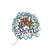 1aoiS S: Starting model for refinement C: citing same article ( |
|---|---|
| Similar structure data |


-Links
 PDBj
PDBj


- Assembly
| Deposited unit | 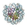
| ||||||||
|---|---|---|---|---|---|---|---|---|---|
| 1 |
| ||||||||
| Unit cell |
|
-Components
-Protein , 4 types, 8 molecules AEBFCGDH
| #1: Protein | Mass: 15303.930 Da / Num. of mol.: 2 Source method: isolated from a genetically manipulated source Source: (gene. exp.)  #2: Protein | Mass: 11323.350 Da / Num. of mol.: 2 Source method: isolated from a genetically manipulated source Source: (gene. exp.) #3: Protein | Mass: 14008.268 Da / Num. of mol.: 2 Source method: isolated from a genetically manipulated source Source: (gene. exp.) #4: Protein | Mass: 13808.033 Da / Num. of mol.: 2 Source method: isolated from a genetically manipulated source Source: (gene. exp.) Gene: hist2h2bf, TGas058p09.1-001 / Production host: |
|---|
-DNA chain / Non-polymers , 2 types, 789 molecules IJ
| #5: DNA chain | Mass: 45054.844 Da / Num. of mol.: 2 / Source method: obtained synthetically #6: Water | ChemComp-HOH / | Mass: 18.015 Da / Num. of mol.: 787 / Source method: isolated from a natural source / Formula: H2O |
|---|
-Details
| Has protein modification | Y |
|---|
-Experimental details
-Experiment
| Experiment | Method: X-RAY DIFFRACTION / Number of used crystals: 1 |
|---|
- Sample preparation
| Crystal | Density Matthews: 2.64 Å3/Da / Density % sol: 53.4 % | ||||||||||||||||||||
|---|---|---|---|---|---|---|---|---|---|---|---|---|---|---|---|---|---|---|---|---|---|
| Crystal grow | Temperature: 292 K / Method: vapor diffusion, sitting drop / pH: 6 Details: Manganese chloride, Potassium chloride, Potassium cacodylate, pH 6.0, VAPOR DIFFUSION, SITTING DROP, temperature 292K | ||||||||||||||||||||
| Components of the solutions |
|
-Data collection
| Diffraction | Mean temperature: 292 K |
|---|---|
| Diffraction source | Source: SYNCHROTRON / Site: ALS  / Beamline: 4.2.2 / Beamline: 4.2.2 |
| Detector | Detector: CCD / Date: Oct 20, 2006 |
| Radiation | Protocol: SINGLE WAVELENGTH / Monochromatic (M) / Laue (L): M / Scattering type: x-ray |
| Radiation wavelength | Relative weight: 1 |
| Reflection | Resolution: 2.2→43 Å / Num. obs: 106458 / % possible obs: 99 % / Observed criterion σ(F): 4.8 / Observed criterion σ(I): 10.2 / Rmerge(I) obs: 0.066 |
| Reflection shell | Resolution: 2.2→2.28 Å / Redundancy: 99 % / Rmerge(I) obs: 0.508 / Mean I/σ(I) obs: 2.6 / % possible all: 99 |
- Processing
| Software |
| ||||||||||||||||
|---|---|---|---|---|---|---|---|---|---|---|---|---|---|---|---|---|---|
| Refinement | Method to determine structure: MOLECULAR REPLACEMENT Starting model: PDB entry 1aoi Resolution: 2.2→43 Å / Cross valid method: throught / Stereochemistry target values: Engh & Huber
| ||||||||||||||||
| Refinement step | Cycle: LAST / Resolution: 2.2→43 Å
| ||||||||||||||||
| Refine LS restraints |
|
