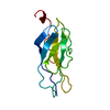
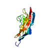
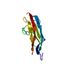
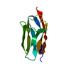
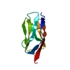
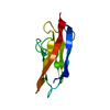
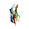
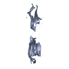
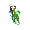
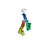
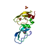
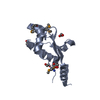
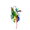
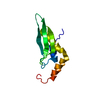
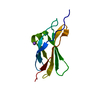
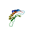
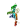
Entry Database : PDB / ID : 2y9rTitle Crystal structure of the M10 domain of Titin TITIN Keywords / / / Function / homology Function Domain/homology Component
/ / / / / / / / / / / / / / / / / / / / / / / / / / / / / / / / / / / / / / / / / / / / / / / / / / / / / / / / / / / / / / / / / / / / / / / / / / / / / / / / / / / / / / / / / / / / / / / / / / Biological species HOMO SAPIENS (human)Method / / / Resolution : 1.9 Å Authors Pernigo, S. / Fukuzawa, A. / Gautel, M. / Steiner, R.A. History Deposition Feb 16, 2011 Deposition site / Processing site Revision 1.0 Mar 2, 2011 Provider / Type Revision 1.1 Jan 25, 2017 Group Atomic model / Data collection ... Atomic model / Data collection / Other / Version format compliance Revision 1.2 Dec 20, 2023 Group Data collection / Database references ... Data collection / Database references / Derived calculations / Other / Refinement description Category chem_comp_atom / chem_comp_bond ... chem_comp_atom / chem_comp_bond / database_2 / pdbx_database_status / pdbx_initial_refinement_model / struct_site Item _database_2.pdbx_DOI / _database_2.pdbx_database_accession ... _database_2.pdbx_DOI / _database_2.pdbx_database_accession / _pdbx_database_status.status_code_sf / _struct_site.pdbx_auth_asym_id / _struct_site.pdbx_auth_comp_id / _struct_site.pdbx_auth_seq_id
Show all Show less
 Movie
Movie Controller


 Open data
 Basic information
 Components
 Keywords
 Function and homology information
 HOMO SAPIENS (human)
 X-RAY DIFFRACTION /
  Authors
 Citation
 Structure visualization
 Downloads & links
 Other downloads


 PDBj
PDBj


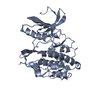
 Assembly

 Mass: 546.646 Da / Num. of mol.: 2 / Source method: obtained synthetically / Formula: C24H50O13 / Comment: precipitant*YM
 Mass: 18.015 Da / Num. of mol.: 44 / Source method: isolated from a natural source / Formula: H2O
 Sample preparation
 / Beamline: I04 / Wavelength: 0.9704
  Processing