 Movie
Movie Controller


+ Open data

- Basic information
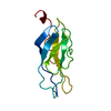
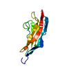
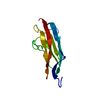
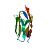
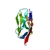
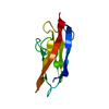
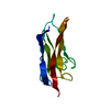
| Entry | Database: PDB / ID: 2wp3 | ||||||
|---|---|---|---|---|---|---|---|
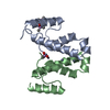
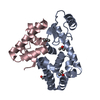
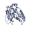
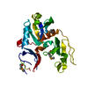
| Title | Crystal structure of the Titin M10-Obscurin like 1 Ig complex | ||||||
 Components Components |
| ||||||
 Keywords Keywords | TRANSFERASE/STRUCTURAL PROTEIN / TRANSFERASE-STRUCTURAL PROTEIN COMPLEX / SARCOMERE / IMMUNOGLOBULIN DOMAIN / LIMB-GIRDLE MUSCULAR DYSTROPHY / SERINE/THREONINE-PROTEIN KINASE / ATP-BINDING / KELCH REPEAT / CARDIOMYOPATHY / CALMODULIN-BINDING | ||||||
| Function / homology |  Function and homology information Function and homology information3M complex / sarcomerogenesis / titin-telethonin complex / structural molecule activity conferring elasticity / skeletal muscle myosin thick filament assembly / telethonin binding / protein localization to Golgi apparatus / detection of muscle stretch / : / muscle alpha-actinin binding ...3M complex / sarcomerogenesis / titin-telethonin complex / structural molecule activity conferring elasticity / skeletal muscle myosin thick filament assembly / telethonin binding / protein localization to Golgi apparatus / detection of muscle stretch / : / muscle alpha-actinin binding / cardiac myofibril assembly / cytoskeletal adaptor activity / cardiac muscle hypertrophy / cardiac muscle tissue morphogenesis / protein kinase regulator activity / mitotic chromosome condensation / positive regulation of dendrite morphogenesis / Striated Muscle Contraction / muscle filament sliding / actinin binding / M band / cardiac muscle cell development / I band / sarcomere organization / structural constituent of muscle / regulation of mitotic nuclear division / Golgi organization / striated muscle thin filament / skeletal muscle thin filament assembly / intercalated disc / striated muscle contraction / skeletal muscle contraction / cardiac muscle contraction / cytoskeleton organization / muscle contraction / condensed nuclear chromosome / positive regulation of protein secretion / response to calcium ion / microtubule cytoskeleton organization / Z disc / actin filament binding / Platelet degranulation / actin cytoskeleton / Neddylation / protein tyrosine kinase activity / protease binding / calmodulin binding / non-specific serine/threonine protein kinase / protein serine kinase activity / protein serine/threonine kinase activity / calcium ion binding / positive regulation of gene expression / centrosome / protein kinase binding / perinuclear region of cytoplasm / enzyme binding / Golgi apparatus / protein homodimerization activity / extracellular exosome / extracellular region / ATP binding / identical protein binding / cytoplasm / cytosol Similarity search - Function | ||||||
| Biological species |  HOMO SAPIENS (human) HOMO SAPIENS (human) | ||||||
| Method |  X-RAY DIFFRACTION / SYNCHROTRON / MOLECULAR REPLACEMENT / Resolution: 1.48 Å X-RAY DIFFRACTION / SYNCHROTRON / MOLECULAR REPLACEMENT / Resolution: 1.48 Å | ||||||
 Authors Authors | Pernigo, S. / Fuzukawa, A. / Gautel, M. / Steiner, R.A. | ||||||
 Citation Citation | Journal: Proc.Natl.Acad.Sci.USA / Year: 2010 Title: Structural Insight Into M-Band Assembly and Mechanics from the Titin-Obscurin-Like-1 Complex. Authors: Pernigo, S. / Fukuzawa, A. / Bertz, M. / Holt, M. / Rief, M. / Steiner, R.A. / Gautel, M. | ||||||
| History |
| ||||||
| Remark 700 | SHEET THE SHEET STRUCTURE OF THIS MOLECULE IS BIFURCATED. IN ORDER TO REPRESENT THIS FEATURE IN ... SHEET THE SHEET STRUCTURE OF THIS MOLECULE IS BIFURCATED. IN ORDER TO REPRESENT THIS FEATURE IN THE SHEET RECORDS BELOW, TWO SHEETS ARE DEFINED. |
- Structure visualization
| Structure viewer | Molecule: MolmilJmol/JSmol |
|---|
- Downloads & links
-Download
| PDBx/mmCIF format | 2wp3.cif.gz | 61.3 KB | Display | PDBx/mmCIF format |
|---|---|---|---|---|
| PDB format | pdb2wp3.ent.gz | 45.2 KB | Display | PDB format |
| PDBx/mmJSON format | 2wp3.json.gz | Tree view | PDBx/mmJSON format | |
| Others |  Other downloads Other downloads |
-Validation report
| Arichive directory | https://data.pdbj.org/pub/pdb/validation_reports/wp/2wp3ftp://data.pdbj.org/pub/pdb/validation_reports/wp/2wp3 | HTTPS FTP |
|---|
-Related structure data


-Links
 PDBj
PDBj


- Assembly
| Deposited unit | 
| ||||||||
|---|---|---|---|---|---|---|---|---|---|
| 1 |
| ||||||||
| Unit cell |
|
-Components
| #1: Protein | Mass: 11125.619 Da / Num. of mol.: 1 / Fragment: IG1, RESIDUES 1-106 Source method: isolated from a genetically manipulated source Source: (gene. exp.) HOMO SAPIENS (human) / Production host:  | ||||||||
|---|---|---|---|---|---|---|---|---|---|
| #2: Protein | Mass: 10981.231 Da / Num. of mol.: 1 / Fragment: M10, RESIDUES 34252-34350 Source method: isolated from a genetically manipulated source Source: (gene. exp.) HOMO SAPIENS (human) / Production host: References: UniProt: Q8WZ42, non-specific serine/threonine protein kinase | ||||||||
| #3: Chemical | ChemComp-GOL /   Mass: 92.094 Da / Num. of mol.: 4 / Source method: obtained synthetically / Formula: C3H8O3 Mass: 92.094 Da / Num. of mol.: 4 / Source method: obtained synthetically / Formula: C3H8O3#4: Chemical | ChemComp-SO4 / |   Mass: 96.063 Da / Num. of mol.: 1 / Source method: obtained synthetically / Formula: SO4 Mass: 96.063 Da / Num. of mol.: 1 / Source method: obtained synthetically / Formula: SO4#5: Water | ChemComp-HOH / |  Mass: 18.015 Da / Num. of mol.: 233 / Source method: isolated from a natural source / Formula: H2O Mass: 18.015 Da / Num. of mol.: 233 / Source method: isolated from a natural source / Formula: H2OHas protein modification | Y | Sequence details | INITIAL GSS ARE FROM THE EXPRESSION | |
-Experimental details
-Experiment
| Experiment | Method: X-RAY DIFFRACTION / Number of used crystals: 1 |
|---|
- Sample preparation
| Crystal | Density Matthews: 2.1 Å3/Da / Density % sol: 41.5 % / Description: NONE |
|---|---|
| Crystal grow | pH: 7 Details: PROTEIN COMPLEX AT 13 MG/ML 2M AMM SULPHATE, 0.1 M BIS-TRIS PH 5.5 |
-Data collection
| Diffraction | Mean temperature: 100 K |
|---|---|
| Diffraction source | Source: SYNCHROTRON / Site: Diamond  / Beamline: I04 / Wavelength: 0.9745 / Beamline: I04 / Wavelength: 0.9745 |
| Detector | Type: ADSC CCD / Detector: CCD / Date: Jan 21, 2009 / Details: MIRRORS |
| Radiation | Monochromator: SI(111) / Protocol: SINGLE WAVELENGTH / Monochromatic (M) / Laue (L): M / Scattering type: x-ray |
| Radiation wavelength | Wavelength: 0.9745 Å / Relative weight: 1 |
| Reflection | Resolution: 1.48→53.38 Å / Num. obs: 30142 / % possible obs: 99.9 % / Observed criterion σ(I): 0 / Redundancy: 5 % / Biso Wilson estimate: 19.38 Å2 / Rmerge(I) obs: 0.06 / Net I/σ(I): 15.9 |
| Reflection shell | Resolution: 1.48→1.56 Å / Redundancy: 3.9 % / Rmerge(I) obs: 0.44 / Mean I/σ(I) obs: 4.6 / % possible all: 99.6 |
- Processing
| Software |
| ||||||||||||||||||||||||||||||||||||||||||||||||||||||||||||||||||||||||||||||||||||||||||||||||||||||||||||||||||||||||||||||||||||||||||||||||||||||||||||||||||||||||||||||||||||||
|---|---|---|---|---|---|---|---|---|---|---|---|---|---|---|---|---|---|---|---|---|---|---|---|---|---|---|---|---|---|---|---|---|---|---|---|---|---|---|---|---|---|---|---|---|---|---|---|---|---|---|---|---|---|---|---|---|---|---|---|---|---|---|---|---|---|---|---|---|---|---|---|---|---|---|---|---|---|---|---|---|---|---|---|---|---|---|---|---|---|---|---|---|---|---|---|---|---|---|---|---|---|---|---|---|---|---|---|---|---|---|---|---|---|---|---|---|---|---|---|---|---|---|---|---|---|---|---|---|---|---|---|---|---|---|---|---|---|---|---|---|---|---|---|---|---|---|---|---|---|---|---|---|---|---|---|---|---|---|---|---|---|---|---|---|---|---|---|---|---|---|---|---|---|---|---|---|---|---|---|---|---|---|---|
| Refinement | Method to determine structure: MOLECULAR REPLACEMENT Starting model: PREVIOUSLY SOLVED STRUCTURE FROM IN-HOUSE DATA Resolution: 1.48→33.19 Å / Cor.coef. Fo:Fc: 0.972 / Cor.coef. Fo:Fc free: 0.953 / SU B: 4.202 / SU ML: 0.07 / TLS residual ADP flag: LIKELY RESIDUAL / Cross valid method: THROUGHOUT / ESU R: 0.079 / ESU R Free: 0.084 / Stereochemistry target values: MAXIMUM LIKELIHOOD Details: HYDROGENS HAVE BEEN ADDED IN THE RIDING POSITIONS. U VALUES RESIDUAL ONLY. ATOM RECORD CONTAINS RESIDUAL B FACTORS ONLY.
| ||||||||||||||||||||||||||||||||||||||||||||||||||||||||||||||||||||||||||||||||||||||||||||||||||||||||||||||||||||||||||||||||||||||||||||||||||||||||||||||||||||||||||||||||||||||
| Solvent computation | Ion probe radii: 0.8 Å / Shrinkage radii: 0.8 Å / VDW probe radii: 1.2 Å / Solvent model: MASK | ||||||||||||||||||||||||||||||||||||||||||||||||||||||||||||||||||||||||||||||||||||||||||||||||||||||||||||||||||||||||||||||||||||||||||||||||||||||||||||||||||||||||||||||||||||||
| Displacement parameters | Biso mean: 12.578 Å2
| ||||||||||||||||||||||||||||||||||||||||||||||||||||||||||||||||||||||||||||||||||||||||||||||||||||||||||||||||||||||||||||||||||||||||||||||||||||||||||||||||||||||||||||||||||||||
| Refinement step | Cycle: LAST / Resolution: 1.48→33.19 Å
| ||||||||||||||||||||||||||||||||||||||||||||||||||||||||||||||||||||||||||||||||||||||||||||||||||||||||||||||||||||||||||||||||||||||||||||||||||||||||||||||||||||||||||||||||||||||
| Refine LS restraints |
|