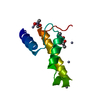
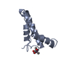
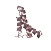
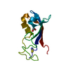
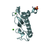
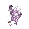
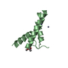
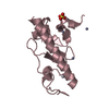
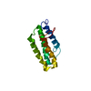
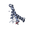
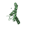
Entry Database : PDB / ID : 2vqhTitle Crystal structure of PorB from Corynebacterium glutamicum (crystal form II) PUTATIVE UNCHARACTERIZED PROTEIN CGL0972 Keywords / / / / / Function / homology Function Domain/homology Component
/ / / / / / Biological species CORYNEBACTERIUM GLUTAMICUM (bacteria)Method / / / Resolution : 2.89 Å Authors Ziegler, K. / Benz, R. / Schulz, G.E. Journal : J.Mol.Biol. / Year : 2008Title : A Putative Alpha-Helical Porin from Corynebacterium Glutamicum.Authors : Ziegler, K. / Benz, R. / Schulz, G.E. History Deposition Mar 15, 2008 Deposition site / Processing site Revision 1.0 May 20, 2008 Provider / Type Revision 1.1 Jun 2, 2011 Group Revision 1.2 Jul 13, 2011 Group Revision 1.3 Oct 16, 2024 Group Data collection / Database references ... Data collection / Database references / Derived calculations / Other / Structure summary Category chem_comp_atom / chem_comp_bond ... chem_comp_atom / chem_comp_bond / citation / database_2 / pdbx_database_status / pdbx_entry_details / pdbx_modification_feature / pdbx_struct_conn_angle / struct_conn Item _citation.page_last / _database_2.pdbx_DOI ... _citation.page_last / _database_2.pdbx_DOI / _database_2.pdbx_database_accession / _pdbx_database_status.status_code_sf / _pdbx_struct_conn_angle.ptnr1_auth_asym_id / _pdbx_struct_conn_angle.ptnr1_auth_comp_id / _pdbx_struct_conn_angle.ptnr1_auth_seq_id / _pdbx_struct_conn_angle.ptnr1_label_asym_id / _pdbx_struct_conn_angle.ptnr1_label_atom_id / _pdbx_struct_conn_angle.ptnr1_label_comp_id / _pdbx_struct_conn_angle.ptnr1_label_seq_id / _pdbx_struct_conn_angle.ptnr1_symmetry / _pdbx_struct_conn_angle.ptnr2_auth_asym_id / _pdbx_struct_conn_angle.ptnr2_auth_seq_id / _pdbx_struct_conn_angle.ptnr2_label_asym_id / _pdbx_struct_conn_angle.ptnr3_auth_asym_id / _pdbx_struct_conn_angle.ptnr3_auth_comp_id / _pdbx_struct_conn_angle.ptnr3_auth_seq_id / _pdbx_struct_conn_angle.ptnr3_label_asym_id / _pdbx_struct_conn_angle.ptnr3_label_atom_id / _pdbx_struct_conn_angle.ptnr3_label_comp_id / _pdbx_struct_conn_angle.ptnr3_label_seq_id / _pdbx_struct_conn_angle.ptnr3_symmetry / _pdbx_struct_conn_angle.value / _struct_conn.pdbx_dist_value / _struct_conn.ptnr1_auth_asym_id / _struct_conn.ptnr1_auth_comp_id / _struct_conn.ptnr1_auth_seq_id / _struct_conn.ptnr1_label_asym_id / _struct_conn.ptnr1_label_atom_id / _struct_conn.ptnr1_label_comp_id / _struct_conn.ptnr1_label_seq_id / _struct_conn.ptnr2_auth_asym_id / _struct_conn.ptnr2_auth_comp_id / _struct_conn.ptnr2_auth_seq_id / _struct_conn.ptnr2_label_asym_id / _struct_conn.ptnr2_label_atom_id / _struct_conn.ptnr2_label_comp_id / _struct_conn.ptnr2_label_seq_id / _struct_conn.ptnr2_symmetry
Show all Show less Remark 650 HELIX DETERMINATION METHOD: AUTHOR PROVIDED.
 Movie
Movie Controller

 Yorodumi
 Open data
 Basic information
 Components
 Keywords
 Function and homology information
 CORYNEBACTERIUM GLUTAMICUM (bacteria)
 X-RAY DIFFRACTION /
  Authors
 Citation
 Structure visualization
 Downloads & links
 Other downloads
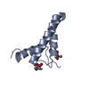

 PDBj
PDBj


 Assembly
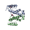

 Mass: 65.409 Da / Num. of mol.: 8 / Source method: obtained synthetically / Formula: Zn
 Mass: 136.989 Da / Num. of mol.: 3 / Source method: obtained synthetically / Formula: C2H6AsO2
 Sample preparation
 / Type:
  Processing