 Movie
Movie Controller

[English] 日本語
 Yorodumi
- PDB-2vql: Crystal structure of PorB from Corynebacterium glutamicum (crysta... -
+ Open data

- Basic information
| Entry | Database: PDB / ID: 2vql | ||||||
|---|---|---|---|---|---|---|---|
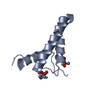
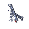
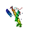
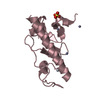
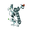
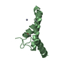
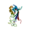
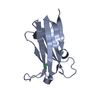
| Title | Crystal structure of PorB from Corynebacterium glutamicum (crystal form III) | ||||||
 Components Components | UNCHARACTERIZED PROTEIN CGL0972 | ||||||
 Keywords Keywords | MEMBRANE PROTEIN / TRANSMEMBRANE / PORIN / MEMBRANE / TRANSPORT / ION TRANSPORT | ||||||
| Function / homology |  Function and homology information Function and homology information | ||||||
| Biological species |  CORYNEBACTERIUM GLUTAMICUM (bacteria) CORYNEBACTERIUM GLUTAMICUM (bacteria) | ||||||
| Method |  X-RAY DIFFRACTION / SYNCHROTRON / MOLECULAR REPLACEMENT / Resolution: 3.16 Å X-RAY DIFFRACTION / SYNCHROTRON / MOLECULAR REPLACEMENT / Resolution: 3.16 Å | ||||||
 Authors Authors | Ziegler, K. / Benz, R. / Schulz, G.E. | ||||||
 Citation Citation | Journal: J.Mol.Biol. / Year: 2008 Title: A Putative Alpha-Helical Porin from Corynebacterium Glutamicum. Authors: Ziegler, K. / Benz, R. / Schulz, G.E. | ||||||
| History |
| ||||||
| Remark 650 | HELIX DETERMINATION METHOD: AUTHOR PROVIDED. |
- Structure visualization
| Structure viewer | Molecule: MolmilJmol/JSmol |
|---|
- Downloads & links
-Download
| PDBx/mmCIF format | 2vql.cif.gz | 128 KB | Display | PDBx/mmCIF format |
|---|---|---|---|---|
| PDB format | pdb2vql.ent.gz | 102.3 KB | Display | PDB format |
| PDBx/mmJSON format | 2vql.json.gz | Tree view | PDBx/mmJSON format | |
| Others |  Other downloads Other downloads |
-Validation report
| Arichive directory | https://data.pdbj.org/pub/pdb/validation_reports/vq/2vqlftp://data.pdbj.org/pub/pdb/validation_reports/vq/2vql | HTTPS FTP |
|---|
-Related structure data


-Links
 PDBj
PDBj


- Assembly
| Deposited unit | 
| ||||||||
|---|---|---|---|---|---|---|---|---|---|
| 1 | 
| ||||||||
| 2 | 
| ||||||||
| 3 | 
| ||||||||
| 4 | 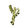
| ||||||||
| Unit cell |
|
-Components
| #1: Protein | Mass: 10643.550 Da / Num. of mol.: 4 / Fragment: RESIDUES 28-126 Source method: isolated from a genetically manipulated source Source: (gene. exp.) CORYNEBACTERIUM GLUTAMICUM (bacteria) / Production host: #2: Chemical | ChemComp-ZN /   Mass: 65.409 Da / Num. of mol.: 12 / Source method: obtained synthetically / Formula: Zn Mass: 65.409 Da / Num. of mol.: 12 / Source method: obtained synthetically / Formula: Zn#3: Chemical | ChemComp-CAC /   Mass: 136.989 Da / Num. of mol.: 4 / Source method: obtained synthetically / Formula: C2H6AsO2 Mass: 136.989 Da / Num. of mol.: 4 / Source method: obtained synthetically / Formula: C2H6AsO2Has protein modification | Y | |
|---|
-Experimental details
-Experiment
| Experiment | Method: X-RAY DIFFRACTION |
|---|
- Sample preparation
| Crystal | Density Matthews: 3.37 Å3/Da / Density % sol: 63.57 % / Description: NONE |
|---|
-Data collection
| Diffraction | Mean temperature: 100 K |
|---|---|
| Diffraction source | Source: SYNCHROTRON / Site: SLS  / Type: SLS / Wavelength: 0.98 / Type: SLS / Wavelength: 0.98 |
| Radiation | Protocol: SINGLE WAVELENGTH / Monochromatic (M) / Laue (L): M / Scattering type: x-ray |
| Radiation wavelength | Wavelength: 0.98 Å / Relative weight: 1 |
| Reflection | Resolution: 3.16→60.75 Å / Num. obs: 10828 / % possible obs: 99.4 % / Observed criterion σ(I): 3 / Redundancy: 8.4 % / Rsym value: 0.06 / Net I/σ(I): 23.9 |
| Reflection shell | Redundancy: 8.8 % / Mean I/σ(I) obs: 6.5 / Rsym value: 0.36 / % possible all: 100 |
- Processing
| Software | Name: REFMAC / Version: 5.2.0019 / Classification: refinement | ||||||||||||||||||||||||||||||||||||||||||||||||||||||||||||||||||||||||||||||||||||||||||||||||||||||||||||||||||||||||||||||||||||||||||||||||||||||||||||||||||||||||||||||||||||||
|---|---|---|---|---|---|---|---|---|---|---|---|---|---|---|---|---|---|---|---|---|---|---|---|---|---|---|---|---|---|---|---|---|---|---|---|---|---|---|---|---|---|---|---|---|---|---|---|---|---|---|---|---|---|---|---|---|---|---|---|---|---|---|---|---|---|---|---|---|---|---|---|---|---|---|---|---|---|---|---|---|---|---|---|---|---|---|---|---|---|---|---|---|---|---|---|---|---|---|---|---|---|---|---|---|---|---|---|---|---|---|---|---|---|---|---|---|---|---|---|---|---|---|---|---|---|---|---|---|---|---|---|---|---|---|---|---|---|---|---|---|---|---|---|---|---|---|---|---|---|---|---|---|---|---|---|---|---|---|---|---|---|---|---|---|---|---|---|---|---|---|---|---|---|---|---|---|---|---|---|---|---|---|---|
| Refinement | Method to determine structure: MOLECULAR REPLACEMENT / Resolution: 3.16→60.75 Å / Cor.coef. Fo:Fc: 0.898 / Cor.coef. Fo:Fc free: 0.87 / SU B: 39.254 / SU ML: 0.392 / Cross valid method: THROUGHOUT / ESU R: 1.225 / ESU R Free: 0.463 / Stereochemistry target values: MAXIMUM LIKELIHOOD / Details: HYDROGENS HAVE BEEN ADDED IN THE RIDING POSITIONS.
| ||||||||||||||||||||||||||||||||||||||||||||||||||||||||||||||||||||||||||||||||||||||||||||||||||||||||||||||||||||||||||||||||||||||||||||||||||||||||||||||||||||||||||||||||||||||
| Solvent computation | Ion probe radii: 0.8 Å / Shrinkage radii: 0.8 Å / VDW probe radii: 1.4 Å / Solvent model: MASK | ||||||||||||||||||||||||||||||||||||||||||||||||||||||||||||||||||||||||||||||||||||||||||||||||||||||||||||||||||||||||||||||||||||||||||||||||||||||||||||||||||||||||||||||||||||||
| Displacement parameters | Biso mean: 109.56 Å2
| ||||||||||||||||||||||||||||||||||||||||||||||||||||||||||||||||||||||||||||||||||||||||||||||||||||||||||||||||||||||||||||||||||||||||||||||||||||||||||||||||||||||||||||||||||||||
| Refinement step | Cycle: LAST / Resolution: 3.16→60.75 Å
| ||||||||||||||||||||||||||||||||||||||||||||||||||||||||||||||||||||||||||||||||||||||||||||||||||||||||||||||||||||||||||||||||||||||||||||||||||||||||||||||||||||||||||||||||||||||
| Refine LS restraints |
|