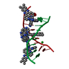
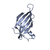
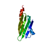
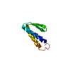
Mass: 12476.979 Da / Num. of mol.: 1 / Fragment: 71 TH DOMAIN FROM THE A-BAND / Mutation: I9V Source method: isolated from a genetically manipulated source Source: (gene. exp.) Homo sapiens (human) / Tissue: MUSCLE / Organ: HEART / Organelle: SARCOMERE / Plasmid: PET8C / Gene (production host): TITIN GENE / Production host: Escherichia coli (E. coli) / Strain (production host): BL21 PLYSS / References: UniProt: Q10466, UniProt: Q8WZ42*PLUS
-
Experimental details
-
Experiment
Experiment
Method: SOLUTION NMR
NMR experiment
Conditions-ID
Experiment-ID
Solution-ID
Type
1
1
1
NOESY
1
2
1
TOCSY
1
3
1
HSQC
1
4
1
3D NOESY-HSQC
NMR details
Text: REPRESENTATIVE MODEL NUMBER: 1 THE STRUCTURE WAS DETERMINED USING A 3D N15-EDITED NOESY-HSQC AND HOMONUCLEAR 2D NOESY EXPERIMENTS.
-
Sample preparation
Details
Contents: 10 MM ACETATE BUFFER
Sample conditions
Ionic strength: 50 mM NACL / pH: 5.6 / Temperature: 310 K
Crystal grow
*PLUS
Method: other / Details: NMR
-
NMR measurement
NMR spectrometer
Type: Bruker AMX600 / Manufacturer: Bruker / Model: AMX600 / Field strength: 600 MHz
-
Processing
Software
Name
Classification
X-PLOR
modelbuilding
X-PLOR
refinement
X-PLOR
phasing
NMR software
Name
Version
Developer
Classification
XPLOR 3.851 (EXTENDED VERSION)
VERSION)
BRUNGER, NILGES
refinement
XPLOR (EXTENDED VERSION)
VERSION)
structuresolution
Refinement
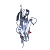
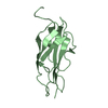
Method: simulated annealing / Software ordinal: 1 Details: STRUCTURE CALCULATIONS WERE PERFORMED WITH THE SUPPORT OF THE ARIA (AMBIGUOUS RESTRAINTS FOR ITERATIVE ASSIGNMENT) PROTOCOL (NILGES ET AL., J. MOL. BIOL. 269, 408-422). 100 STRUCTURES WERE ...Details: STRUCTURE CALCULATIONS WERE PERFORMED WITH THE SUPPORT OF THE ARIA (AMBIGUOUS RESTRAINTS FOR ITERATIVE ASSIGNMENT) PROTOCOL (NILGES ET AL., J. MOL. BIOL. 269, 408-422). 100 STRUCTURES WERE CALCULATED OF WHICH THE BEST 50 STRUCTURES WERE SELECTED.
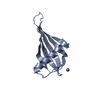
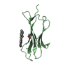
NMR ensemble
Conformer selection criteria: LEAST RESTRAINT VIOLATION / Conformers calculated total number: 1 / Conformers submitted total number: 50
+
About Yorodumi
-
News
-
Feb 9, 2022. New format data for meta-information of EMDB entries
New format data for meta-information of EMDB entries
Version 3 of the EMDB header file is now the official format.
The previous official version 1.9 will be removed from the archive.
In the structure databanks used in Yorodumi, some data are registered as the other names, "COVID-19 virus" and "2019-nCoV". Here are the details of the virus and the list of structure data.
Jan 31, 2019. EMDB accession codes are about to change! (news from PDBe EMDB page)
EMDB accession codes are about to change! (news from PDBe EMDB page)
The allocation of 4 digits for EMDB accession codes will soon come to an end. Whilst these codes will remain in use, new EMDB accession codes will include an additional digit and will expand incrementally as the available range of codes is exhausted. The current 4-digit format prefixed with “EMD-” (i.e. EMD-XXXX) will advance to a 5-digit format (i.e. EMD-XXXXX), and so on. It is currently estimated that the 4-digit codes will be depleted around Spring 2019, at which point the 5-digit format will come into force.
The EM Navigator/Yorodumi systems omit the EMD- prefix.
Related info.:Q: What is EMD? / ID/Accession-code notation in Yorodumi/EM Navigator
Yorodumi is a browser for structure data from EMDB, PDB, SASBDB, etc.
This page is also the successor to EM Navigator detail page, and also detail information page/front-end page for Omokage search.
The word "yorodu" (or yorozu) is an old Japanese word meaning "ten thousand". "mi" (miru) is to see.
Related info.:EMDB / PDB / SASBDB / Comparison of 3 databanks / Yorodumi Search / Aug 31, 2016. New EM Navigator & Yorodumi / Yorodumi Papers / Jmol/JSmol / Function and homology information / Changes in new EM Navigator and Yorodumi
 Movie
Movie Controller


 Open data
 Basic information
 Components
 Keywords
 Function and homology information
 Homo sapiens (human)
 Authors
 Citation
 Structure visualization
 Downloads & links
 Other downloads
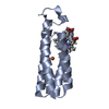

 PDBj
PDBj


 Assembly

 HSQC
HSQC Sample preparation
 Processing