 Movie
Movie Controller


+ Open data

- Basic information
| Entry | Database: PDB / ID: 1i9e | ||||||
|---|---|---|---|---|---|---|---|
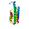
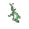
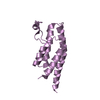
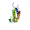
| Title | TCR DOMAIN | ||||||
 Components Components | CYTOTOXIC TCELL VALPHA DOMAIN | ||||||
 Keywords Keywords | IMMUNE SYSTEM / Ig-like domain / T Cell receptor | ||||||
| Function / homology |  Function and homology information Function and homology information | ||||||
| Biological species |  | ||||||
| Method |  X-RAY DIFFRACTION / SYNCHROTRON / MOLECULAR REPLACEMENT / Resolution: 2.5 Å X-RAY DIFFRACTION / SYNCHROTRON / MOLECULAR REPLACEMENT / Resolution: 2.5 Å | ||||||
 Authors Authors | Rudolph, M.G. / Huang, M. / Teyton, L. / Wilson, I.A. | ||||||
 Citation Citation | Journal: J.Mol.Biol. / Year: 2001 Title: Crystal structure of an isolated V(alpha) domain of the 2C T-cell receptor. Authors: Rudolph, M.G. / Huang, M. / Teyton, L. / Wilson, I.A. | ||||||
| History |
|
- Structure visualization
| Structure viewer | Molecule: MolmilJmol/JSmol |
|---|
- Downloads & links
-Download
| PDBx/mmCIF format | 1i9e.cif.gz | 36.9 KB | Display | PDBx/mmCIF format |
|---|---|---|---|---|
| PDB format | pdb1i9e.ent.gz | 25.3 KB | Display | PDB format |
| PDBx/mmJSON format | 1i9e.json.gz | Tree view | PDBx/mmJSON format | |
| Others |  Other downloads Other downloads |
-Validation report
| Arichive directory | https://data.pdbj.org/pub/pdb/validation_reports/i9/1i9eftp://data.pdbj.org/pub/pdb/validation_reports/i9/1i9e | HTTPS FTP |
|---|
-Related structure data
| Related structure data |  1tcrS S: Starting model for refinement |
|---|---|
| Similar structure data |


-Links
 PDBj
PDBj

- Assembly
| Deposited unit | 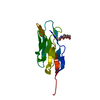
| ||||||||
|---|---|---|---|---|---|---|---|---|---|
| 1 |
| ||||||||
| Unit cell |
|
-Components
| #1: Protein | Mass: 12774.330 Da / Num. of mol.: 1 / Fragment: N-TERMINAL DOMAIN OF ALPHA CHAIN Source method: isolated from a genetically manipulated source Source: (gene. exp.)  |
|---|---|
| #2: Sugar | ChemComp-NAG /   Type: D-saccharide, beta linking / Mass: 221.208 Da / Num. of mol.: 1 Type: D-saccharide, beta linking / Mass: 221.208 Da / Num. of mol.: 1Source method: isolated from a genetically manipulated source Formula: C8H15NO6 |
| #3: Water | ChemComp-HOH /  Mass: 18.015 Da / Num. of mol.: 44 / Source method: isolated from a natural source / Formula: H2O Mass: 18.015 Da / Num. of mol.: 44 / Source method: isolated from a natural source / Formula: H2O |
| Has protein modification | Y |
-Experimental details
-Experiment
| Experiment | Method: X-RAY DIFFRACTION / Number of used crystals: 1 |
|---|
- Sample preparation
| Crystal | Density Matthews: 3.34 Å3/Da / Density % sol: 63.17 % | |||||||||||||||||||||||||||||||||||
|---|---|---|---|---|---|---|---|---|---|---|---|---|---|---|---|---|---|---|---|---|---|---|---|---|---|---|---|---|---|---|---|---|---|---|---|---|
| Crystal grow | Temperature: 295 K / Method: vapor diffusion / pH: 7.5 Details: di-sodium tartrate, NaCl, imidazole/HCl, pH 7.5, VAPOR DIFFUSION, temperature 295K | |||||||||||||||||||||||||||||||||||
| Crystal grow | *PLUS Temperature: 25 ℃ / Method: vapor diffusion, sitting drop | |||||||||||||||||||||||||||||||||||
| Components of the solutions | *PLUS
|
-Data collection
| Diffraction | Mean temperature: 85 K |
|---|---|
| Diffraction source | Source: SYNCHROTRON / Site: SSRL  / Beamline: BL9-1 / Wavelength: 0.783 Å / Beamline: BL9-1 / Wavelength: 0.783 Å |
| Detector | Type: MARRESEARCH / Detector: IMAGE PLATE |
| Radiation | Protocol: SINGLE WAVELENGTH / Monochromatic (M) / Laue (L): M / Scattering type: x-ray |
| Radiation wavelength | Wavelength: 0.783 Å / Relative weight: 1 |
| Reflection | Resolution: 2.5→37.5 Å / Num. obs: 6370 / % possible obs: 99.6 % / Observed criterion σ(I): 1 / Redundancy: 10.2 % / Biso Wilson estimate: 47 Å2 / Rmerge(I) obs: 0.114 / Net I/σ(I): 22.2 |
| Reflection shell | Resolution: 2.5→2.54 Å / Redundancy: 10.7 % / Rmerge(I) obs: 0.748 / Mean I/σ(I) obs: 3.4 / Num. unique all: 306 / % possible all: 100 |
| Reflection | *PLUS Num. obs: 6260 |
| Reflection shell | *PLUS Highest resolution: 2.5 Å / % possible obs: 100 % / Num. unique obs: 306 |
- Processing
| Software |
| ||||||||||||||||||||||||||||||||||||||||
|---|---|---|---|---|---|---|---|---|---|---|---|---|---|---|---|---|---|---|---|---|---|---|---|---|---|---|---|---|---|---|---|---|---|---|---|---|---|---|---|---|---|
| Refinement | Method to determine structure: MOLECULAR REPLACEMENT Starting model: Valpha domain of 2C TCR (PDB entry 1tcr) residues 1-115 Resolution: 2.5→37.5 Å / SU B: 12.635 / SU ML: 0.288 / TLS residual ADP flag: LIKELY RESIDUAL / Isotropic thermal model: isotropic / Cross valid method: THROUGHOUT / σ(F): 0 / σ(I): 0 / ESU R: 0.262 / ESU R Free: 0.262 / Stereochemistry target values: Engh & Huber Details: HYDROGENS ADDED IN THE RIDING POSITIONS DURING REFINEMENT AND HAVE NOT BEEN OUTPUT AS COORDINATES.
| ||||||||||||||||||||||||||||||||||||||||
| Displacement parameters | Biso mean: 25.866 Å2
| ||||||||||||||||||||||||||||||||||||||||
| Refinement step | Cycle: LAST / Resolution: 2.5→37.5 Å
| ||||||||||||||||||||||||||||||||||||||||
| Refine LS restraints |
| ||||||||||||||||||||||||||||||||||||||||
| LS refinement shell | Highest resolution: 2.5 Å / Rfactor Rfree: 0.3 / Rfactor Rwork: 0.2 | ||||||||||||||||||||||||||||||||||||||||
| Refinement TLS params. | Method: refined / Origin x: 97.465 Å / Origin y: 68.107 Å / Origin z: 31.935 Å
| ||||||||||||||||||||||||||||||||||||||||
| Software | *PLUS Name: REFMAC / Version: 5 / Classification: refinement | ||||||||||||||||||||||||||||||||||||||||
| Refinement | *PLUS σ(F): 0 / % reflection Rfree: 10.8 % / Rfactor all: 0.20632 / Rfactor obs: 0.203 | ||||||||||||||||||||||||||||||||||||||||
| Solvent computation | *PLUS | ||||||||||||||||||||||||||||||||||||||||
| Displacement parameters | *PLUS | ||||||||||||||||||||||||||||||||||||||||
| Refine LS restraints | *PLUS
| ||||||||||||||||||||||||||||||||||||||||
| LS refinement shell | *PLUS Highest resolution: 2.5 Å / Lowest resolution: 2.57 Å / Rfactor Rfree: 0.339 / Rfactor obs: 0.242 |
