 Movie
Movie Controller


+ Open data

- Basic information
| Entry | Database: PDB / ID: 2vvw | ||||||
|---|---|---|---|---|---|---|---|
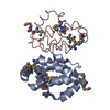
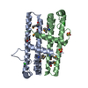
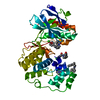
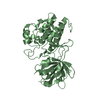
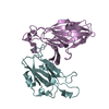
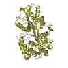
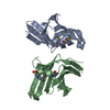
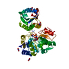
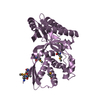
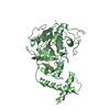
| Title | Structure of Vaccinia virus protein A52 | ||||||
 Components Components | PROTEIN A52 | ||||||
 Keywords Keywords | VIRAL PROTEIN / IRAK2 / TRAF6 / BCL-2 FAMILY / VACCINIA VIRUS / IMMUNOMODULATOR / NF-KB ACTIVATION / HOST-VIRUS INTERACTION | ||||||
| Function / homology |  Function and homology information Function and homology informationsymbiont-mediated suppression of host TRAF-mediated signal transduction / protein sequestering activity / symbiont-mediated suppression of host NF-kappaB cascade / host cell cytoplasm / symbiont-mediated suppression of host innate immune response Similarity search - Function | ||||||
| Biological species |  VACCINIA VIRUS VACCINIA VIRUS | ||||||
| Method |  X-RAY DIFFRACTION / SYNCHROTRON / MAD / Resolution: 1.9 Å X-RAY DIFFRACTION / SYNCHROTRON / MAD / Resolution: 1.9 Å | ||||||
 Authors Authors | Graham, S.C. / Bahar, M.W. / Cooray, S. / Chen, R.A.-J. / Whalen, D.M. / Abrescia, N.G.A. / Alderton, D. / Owens, R.J. / Stuart, D.I. / Smith, G.L. / Grimes, J.M. | ||||||
 Citation Citation | Journal: Plos Pathog. / Year: 2008 Title: Vaccinia Virus Proteins A52 and B14 Share a Bcl-2-Like Fold But Have Evolved to Inhibit NF-kappaB Rather Than Apoptosis Authors: Graham, S.C. / Bahar, M.W. / Cooray, S. / Chen, R.A.-J. / Whalen, D.M. / Abrescia, N.G.A. / Alderton, D. / Owens, R.J. / Stuart, D.I. / Smith, G.L. / Grimes, J.M. | ||||||
| History |
| ||||||
| Remark 650 | HELIX DETERMINATION METHOD: AUTHOR PROVIDED. |
- Structure visualization
| Structure viewer | Molecule: MolmilJmol/JSmol |
|---|
- Downloads & links
-Download
| PDBx/mmCIF format | 2vvw.cif.gz | 139.8 KB | Display | PDBx/mmCIF format |
|---|---|---|---|---|
| PDB format | pdb2vvw.ent.gz | 111.5 KB | Display | PDB format |
| PDBx/mmJSON format | 2vvw.json.gz | Tree view | PDBx/mmJSON format | |
| Others |  Other downloads Other downloads |
-Validation report
| Arichive directory | https://data.pdbj.org/pub/pdb/validation_reports/vv/2vvwftp://data.pdbj.org/pub/pdb/validation_reports/vv/2vvw | HTTPS FTP |
|---|
-Related structure data


-Links
 PDBj
PDBj- Assembly
| Deposited unit | 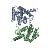
| ||||||||
|---|---|---|---|---|---|---|---|---|---|
| 1 |
| ||||||||
| 2 | 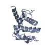
| ||||||||
| 3 | 
| ||||||||
| Unit cell |
|
-Components
| #1: Protein | Mass: 19589.703 Da / Num. of mol.: 2 / Fragment: RESIDUES 37-190 Source method: isolated from a genetically manipulated source Source: (gene. exp.) VACCINIA VIRUS / Strain: WESTERN RESERVE / Plasmid: POPINF / Production host:  #2: Water | ChemComp-HOH / |  Mass: 18.015 Da / Num. of mol.: 207 / Source method: isolated from a natural source / Formula: H2O Mass: 18.015 Da / Num. of mol.: 207 / Source method: isolated from a natural source / Formula: H2OSequence details | RESIDUES 1-36 ARE REMOVED, AN N-TERMINAL START CODON IS ADDED AND A C-TERMINAL LYS-HIS6 ...RESIDUES 1-36 ARE REMOVED, AN N-TERMINAL START CODON IS ADDED AND A C-TERMINAL LYS-HIS6 PURIFICATI | |
|---|
-Experimental details
-Experiment
| Experiment | Method: X-RAY DIFFRACTION / Number of used crystals: 1 |
|---|
- Sample preparation
| Crystal | Density Matthews: 2.55 Å3/Da / Density % sol: 51.84 % / Description: NONE |
|---|---|
| Crystal grow | Temperature: 294 K / Method: vapor diffusion, hanging drop Details: HANGING DROPS CONTAINING 1 UL PROTEIN (5.3 MG/ML IN 20 MM TRIS, 200 MM NACL, PH 7.5) AND 1 UL RESERVOIR SOLUTION (0.2 M TRISODIUM CITRATE, 18% (W/V) PEG 3350) WERE EQUILIBRATED AGAINST 500 ...Details: HANGING DROPS CONTAINING 1 UL PROTEIN (5.3 MG/ML IN 20 MM TRIS, 200 MM NACL, PH 7.5) AND 1 UL RESERVOIR SOLUTION (0.2 M TRISODIUM CITRATE, 18% (W/V) PEG 3350) WERE EQUILIBRATED AGAINST 500 UL RESERVOIRS AT 21 C. CRYSTALS WERE CRYOPROTECTED IN RESERVOIR SOLUTION SUPPLEMENTED WITH 20% (V/V) GLYCEROL. |
-Data collection
| Diffraction | Mean temperature: 100 K |
|---|---|
| Diffraction source | Source: SYNCHROTRON / Site: ESRF  / Beamline: ID14-4 / Wavelength: 0.9395 / Beamline: ID14-4 / Wavelength: 0.9395 |
| Detector | Type: ADSC CCD / Detector: CCD / Date: Aug 27, 2007 / Details: MIRRORS |
| Radiation | Monochromator: SILICON (111) CRYSTAL / Protocol: SINGLE WAVELENGTH / Monochromatic (M) / Laue (L): M / Scattering type: x-ray |
| Radiation wavelength | Wavelength: 0.9395 Å / Relative weight: 1 |
| Reflection | Resolution: 1.9→50 Å / Num. obs: 30971 / % possible obs: 99.9 % / Observed criterion σ(I): -3 / Redundancy: 4.1 % / Biso Wilson estimate: 24.16 Å2 / Rmerge(I) obs: 0.08 / Net I/σ(I): 12.1 |
| Reflection shell | Resolution: 1.9→1.93 Å / Redundancy: 4.1 % / Rmerge(I) obs: 0.71 / Mean I/σ(I) obs: 1.9 / % possible all: 99.7 |
- Processing
| Software |
| ||||||||||||||||||||||||||||||||||||||||||||||||||||||||||||||||||||||||||||||||||||||||||||||||||||||||||||||||||||||||||||||||||||||||||||||||||||||||||||||||||||||||||||||||||||||
|---|---|---|---|---|---|---|---|---|---|---|---|---|---|---|---|---|---|---|---|---|---|---|---|---|---|---|---|---|---|---|---|---|---|---|---|---|---|---|---|---|---|---|---|---|---|---|---|---|---|---|---|---|---|---|---|---|---|---|---|---|---|---|---|---|---|---|---|---|---|---|---|---|---|---|---|---|---|---|---|---|---|---|---|---|---|---|---|---|---|---|---|---|---|---|---|---|---|---|---|---|---|---|---|---|---|---|---|---|---|---|---|---|---|---|---|---|---|---|---|---|---|---|---|---|---|---|---|---|---|---|---|---|---|---|---|---|---|---|---|---|---|---|---|---|---|---|---|---|---|---|---|---|---|---|---|---|---|---|---|---|---|---|---|---|---|---|---|---|---|---|---|---|---|---|---|---|---|---|---|---|---|---|---|
| Refinement | Method to determine structure: MAD Starting model: NONE Resolution: 1.9→33.77 Å / Cor.coef. Fo:Fc: 0.964 / Cor.coef. Fo:Fc free: 0.948 / SU B: 6.892 / SU ML: 0.099 / Cross valid method: THROUGHOUT / ESU R: 0.132 / ESU R Free: 0.127 / Stereochemistry target values: MAXIMUM LIKELIHOOD Details: HYDROGENS HAVE BEEN ADDED IN THE RIDING POSITIONS. B VALUES INCLUDE TLS CONTRIBUTIONS.
| ||||||||||||||||||||||||||||||||||||||||||||||||||||||||||||||||||||||||||||||||||||||||||||||||||||||||||||||||||||||||||||||||||||||||||||||||||||||||||||||||||||||||||||||||||||||
| Solvent computation | Ion probe radii: 0.8 Å / Shrinkage radii: 0.8 Å / VDW probe radii: 1.2 Å / Solvent model: MASK | ||||||||||||||||||||||||||||||||||||||||||||||||||||||||||||||||||||||||||||||||||||||||||||||||||||||||||||||||||||||||||||||||||||||||||||||||||||||||||||||||||||||||||||||||||||||
| Displacement parameters | Biso mean: 26.03 Å2
| ||||||||||||||||||||||||||||||||||||||||||||||||||||||||||||||||||||||||||||||||||||||||||||||||||||||||||||||||||||||||||||||||||||||||||||||||||||||||||||||||||||||||||||||||||||||
| Refinement step | Cycle: LAST / Resolution: 1.9→33.77 Å
| ||||||||||||||||||||||||||||||||||||||||||||||||||||||||||||||||||||||||||||||||||||||||||||||||||||||||||||||||||||||||||||||||||||||||||||||||||||||||||||||||||||||||||||||||||||||
| Refine LS restraints |
|