 Movie
Movie Controller


+ Open data

- Basic information
| Entry | Database: PDB / ID: 2i39 | ||||||
|---|---|---|---|---|---|---|---|
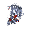
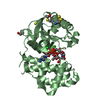
| Title | Crystal structure of Vaccinia virus N1L protein | ||||||
 Components Components | Protein N1 | ||||||
 Keywords Keywords | VIRAL PROTEIN / All Alpha | ||||||
| Function / homology |  Function and homology information Function and homology informationsymbiont-mediated perturbation of host apoptosis / symbiont-mediated perturbation of host defense response / symbiont-mediated suppression of host NF-kappaB cascade Similarity search - Function | ||||||
| Biological species |  Vaccinia virus Vaccinia virus | ||||||
| Method |  X-RAY DIFFRACTION / SYNCHROTRON / SAD / Resolution: 2.2 Å X-RAY DIFFRACTION / SYNCHROTRON / SAD / Resolution: 2.2 Å | ||||||
 Authors Authors | Aoyagi, M. / Aleshin, A.E. / Stec, B. / Liddington, R.C. | ||||||
 Citation Citation | Journal: Protein Sci. / Year: 2007 Title: Vaccinia virus N1L protein resembles a B cell lymphoma-2 (Bcl-2) family protein. Authors: Aoyagi, M. / Zhai, D. / Jin, C. / Aleshin, A.E. / Stec, B. / Reed, J.C. / Liddington, R.C. | ||||||
| History |
|
- Structure visualization
| Structure viewer | Molecule: MolmilJmol/JSmol |
|---|
- Downloads & links
-Download
| PDBx/mmCIF format | 2i39.cif.gz | 158.9 KB | Display | PDBx/mmCIF format |
|---|---|---|---|---|
| PDB format | pdb2i39.ent.gz | 127.1 KB | Display | PDB format |
| PDBx/mmJSON format | 2i39.json.gz | Tree view | PDBx/mmJSON format | |
| Others |  Other downloads Other downloads |
-Validation report
| Arichive directory | https://data.pdbj.org/pub/pdb/validation_reports/i3/2i39ftp://data.pdbj.org/pub/pdb/validation_reports/i3/2i39 | HTTPS FTP |
|---|
-Related structure data
| Similar structure data |
|---|


-Links
 PDBj
PDBj- Assembly
| Deposited unit | 
| ||||||||
|---|---|---|---|---|---|---|---|---|---|
| 1 | 
| ||||||||
| 2 | 
| ||||||||
| 3 | 
| ||||||||
| Unit cell |
| ||||||||
| Details | The asymmetric unit contains three dimers (A/B, C/D & E/F), which likely represent the biological assembly. |
-Components
| #1: Protein | Mass: 16149.409 Da / Num. of mol.: 6 Source method: isolated from a genetically manipulated source Source: (gene. exp.) Vaccinia virus / Genus: Orthopoxvirus / Strain: Western Reserve / Gene: N1L (VACWR028) / Plasmid: pET15b / Production host:  #2: Chemical |   Mass: 118.174 Da / Num. of mol.: 2 / Source method: obtained synthetically / Formula: C6H14O2 / Comment: precipitant*YM Mass: 118.174 Da / Num. of mol.: 2 / Source method: obtained synthetically / Formula: C6H14O2 / Comment: precipitant*YM#3: Water | ChemComp-HOH / |  Mass: 18.015 Da / Num. of mol.: 282 / Source method: isolated from a natural source / Formula: H2O Mass: 18.015 Da / Num. of mol.: 282 / Source method: isolated from a natural source / Formula: H2O |
|---|
-Experimental details
-Experiment
| Experiment | Method: X-RAY DIFFRACTION / Number of used crystals: 1 |
|---|
- Sample preparation
| Crystal | Density Matthews: 2.53 Å3/Da / Density % sol: 51.41 % |
|---|---|
| Crystal grow | Temperature: 290 K / Method: vapor diffusion, hanging drop / pH: 8 Details: 10% PEG 4K, 0.1 M Tris HCl, 0.1 M sodium-potassium tartrate, pH 8, VAPOR DIFFUSION, HANGING DROP, temperature 290K |
-Data collection
| Diffraction | Mean temperature: 100 K |
|---|---|
| Diffraction source | Source: SYNCHROTRON / Site: ALS  / Beamline: 12.3.1 / Wavelength: 1.12 Å / Beamline: 12.3.1 / Wavelength: 1.12 Å |
| Detector | Type: ADSC QUANTUM 315 / Detector: CCD / Date: Dec 17, 2005 |
| Radiation | Monochromator: Si 111 Channel / Protocol: SINGLE WAVELENGTH / Monochromatic (M) / Laue (L): M / Scattering type: x-ray |
| Radiation wavelength | Wavelength: 1.12 Å / Relative weight: 1 |
| Reflection | Resolution: 2.2→30 Å / Num. all: 49469 / Num. obs: 47333 / % possible obs: 95.7 % / Observed criterion σ(I): 4 / Redundancy: 2.8 % / Biso Wilson estimate: 27.2 Å2 / Rsym value: 0.05 / Net I/σ(I): 12.7 |
| Reflection shell | Resolution: 2.2→2.28 Å / Redundancy: 1.9 % / Mean I/σ(I) obs: 2.1 / Num. unique all: 3874 / Rsym value: 0.282 / % possible all: 79.3 |
- Processing
| Software |
| ||||||||||||||||||||||||||||||||||||
|---|---|---|---|---|---|---|---|---|---|---|---|---|---|---|---|---|---|---|---|---|---|---|---|---|---|---|---|---|---|---|---|---|---|---|---|---|---|
| Refinement | Method to determine structure: SAD / Resolution: 2.2→30 Å / Rfactor Rfree error: 0.005 / Data cutoff high absF: 1498118.07 / Data cutoff low absF: 0 / Isotropic thermal model: RESTRAINED / Cross valid method: THROUGHOUT / σ(F): 0 / Stereochemistry target values: Engh & Huber
| ||||||||||||||||||||||||||||||||||||
| Solvent computation | Solvent model: FLAT MODEL / Bsol: 31.3715 Å2 / ksol: 0.304499 e/Å3 | ||||||||||||||||||||||||||||||||||||
| Displacement parameters | Biso mean: 40.864 Å2
| ||||||||||||||||||||||||||||||||||||
| Refine analyze |
| ||||||||||||||||||||||||||||||||||||
| Refinement step | Cycle: LAST / Resolution: 2.2→30 Å
| ||||||||||||||||||||||||||||||||||||
| Refine LS restraints |
| ||||||||||||||||||||||||||||||||||||
| LS refinement shell | Resolution: 2.2→2.34 Å / Rfactor Rfree error: 0.017 / Total num. of bins used: 6
| ||||||||||||||||||||||||||||||||||||
| Xplor file |
|
