 Movie
Movie Controller

[English] 日本語
 Yorodumi
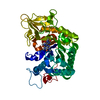
- PDB-2jbx: Crystal Structure of the myxoma virus anti-apoptotic protein M11L -
+ Open data

- Basic information
| Entry | Database: PDB / ID: 2jbx | ||||||
|---|---|---|---|---|---|---|---|
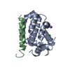
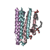
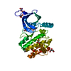
| Title | Crystal Structure of the myxoma virus anti-apoptotic protein M11L | ||||||
 Components Components | M11L PROTEIN | ||||||
 Keywords Keywords | APOPTOSIS / BCL-2 FAMILY / MYXOMA VIRUS | ||||||
| Function / homology | Poxvirus F1/C10 / Apoptosis regulator M11L like / symbiont-mediated suppression of host apoptosis / Blc2-like / Apoptosis Regulator Bcl-x / Bcl-2-like superfamily / Orthogonal Bundle / Mainly Alpha / Apoptosis regulator M11L Function and homology information Function and homology information | ||||||
| Biological species |  MYXOMA VIRUS MYXOMA VIRUS | ||||||
| Method |  X-RAY DIFFRACTION / MOLECULAR REPLACEMENT / Resolution: 2.73 Å X-RAY DIFFRACTION / MOLECULAR REPLACEMENT / Resolution: 2.73 Å | ||||||
 Authors Authors | Kvansakul, M. / Van Delft, M.F. / Lee, E.F. / Gulbis, J.M. / Fairlie, W.D. / Huang, D.C.S. / Colman, P.M. | ||||||
 Citation Citation | Journal: Mol.Cell / Year: 2007 Title: A Structural Viral Mimic of Prosurvival Bcl-2: A Pivotal Role for Sequestering Proapoptotic Bax and Bak. Authors: Kvansakul, M. / Van Delft, M.F. / Lee, E.F. / Gulbis, J.M. / Fairlie, W.D. / Huang, D.C.S. / Colman, P.M. | ||||||
| History |
|
- Structure visualization
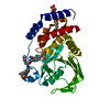
| Structure viewer | Molecule: MolmilJmol/JSmol |
|---|
- Downloads & links
-Download
| PDBx/mmCIF format | 2jbx.cif.gz | 65.9 KB | Display | PDBx/mmCIF format |
|---|---|---|---|---|
| PDB format | pdb2jbx.ent.gz | 49.8 KB | Display | PDB format |
| PDBx/mmJSON format | 2jbx.json.gz | Tree view | PDBx/mmJSON format | |
| Others |  Other downloads Other downloads |
-Validation report
| Arichive directory | https://data.pdbj.org/pub/pdb/validation_reports/jb/2jbxftp://data.pdbj.org/pub/pdb/validation_reports/jb/2jbx | HTTPS FTP |
|---|
-Related structure data


-Links
 PDBj
PDBj
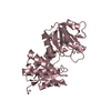
- Assembly
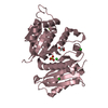
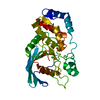
| Deposited unit | 
| ||||||||
|---|---|---|---|---|---|---|---|---|---|
| 1 |
| ||||||||
| Unit cell |
|
-Components
| #1: Protein | Mass: 16881.328 Da / Num. of mol.: 2 / Fragment: RESIDUES 1-132 Source method: isolated from a genetically manipulated source Source: (gene. exp.) MYXOMA VIRUS / Strain: LAUSANNE / Production host:  #2: Water | ChemComp-HOH / |  Mass: 18.015 Da / Num. of mol.: 67 / Source method: isolated from a natural source / Formula: H2O Mass: 18.015 Da / Num. of mol.: 67 / Source method: isolated from a natural source / Formula: H2OHas protein modification | Y | |
|---|
-Experimental details
-Experiment
| Experiment | Method: X-RAY DIFFRACTION / Number of used crystals: 1 |
|---|
- Sample preparation
| Crystal | Density Matthews: 2.19 Å3/Da / Density % sol: 43.82 % / Description: PDB ENTRY FOR M11L BAK COMPLEX TO BE SUBMITTED |
|---|---|
| Crystal grow | pH: 7.2 Details: 1.8 M AMMONIUM SULFATE, 10 MM KSCN, 100 MM HEPES PH 7.2 |
-Data collection
| Diffraction | Mean temperature: 100 K |
|---|---|
| Diffraction source | Source: ROTATING ANODE / Type: RIGAKU MICROMAX-007 / Wavelength: 1.5418 |
| Detector | Type: RIGAKU IMAGE PLATE / Detector: IMAGE PLATE / Date: Dec 7, 2005 / Details: AXCO CAPILLARY OPTICS |
| Radiation | Monochromator: NI FILTER / Protocol: SINGLE WAVELENGTH / Monochromatic (M) / Laue (L): M / Scattering type: x-ray |
| Radiation wavelength | Wavelength: 1.5418 Å / Relative weight: 1 |
| Reflection | Resolution: 2.73→20 Å / Num. obs: 7483 / % possible obs: 97.3 % / Observed criterion σ(I): 3 / Redundancy: 2.7 % / Rmerge(I) obs: 0.11 / Net I/σ(I): 9 |
| Reflection shell | Resolution: 2.73→2.83 Å / Redundancy: 2.4 % / Rmerge(I) obs: 0.35 / Mean I/σ(I) obs: 3 / % possible all: 91.6 |
- Processing
| Software |
| ||||||||||||||||||||||||||||||||||||||||||||||||||||||||||||||||||||||||||||||||||||||||||||||||||||||||||||||||||||||||||||||||||||||||||||||||||||||||||||||||||||||||||||||||||||||
|---|---|---|---|---|---|---|---|---|---|---|---|---|---|---|---|---|---|---|---|---|---|---|---|---|---|---|---|---|---|---|---|---|---|---|---|---|---|---|---|---|---|---|---|---|---|---|---|---|---|---|---|---|---|---|---|---|---|---|---|---|---|---|---|---|---|---|---|---|---|---|---|---|---|---|---|---|---|---|---|---|---|---|---|---|---|---|---|---|---|---|---|---|---|---|---|---|---|---|---|---|---|---|---|---|---|---|---|---|---|---|---|---|---|---|---|---|---|---|---|---|---|---|---|---|---|---|---|---|---|---|---|---|---|---|---|---|---|---|---|---|---|---|---|---|---|---|---|---|---|---|---|---|---|---|---|---|---|---|---|---|---|---|---|---|---|---|---|---|---|---|---|---|---|---|---|---|---|---|---|---|---|---|---|
| Refinement | Method to determine structure: MOLECULAR REPLACEMENT Starting model: M11L BAK COMPLEX Resolution: 2.73→20 Å / Cor.coef. Fo:Fc: 0.932 / Cor.coef. Fo:Fc free: 0.839 / SU B: 18.914 / SU ML: 0.387 / Cross valid method: THROUGHOUT / ESU R Free: 0.445 / Stereochemistry target values: MAXIMUM LIKELIHOOD / Details: HYDROGENS HAVE BEEN ADDED IN THE RIDING POSITIONS.
| ||||||||||||||||||||||||||||||||||||||||||||||||||||||||||||||||||||||||||||||||||||||||||||||||||||||||||||||||||||||||||||||||||||||||||||||||||||||||||||||||||||||||||||||||||||||
| Solvent computation | Ion probe radii: 0.8 Å / Shrinkage radii: 0.8 Å / VDW probe radii: 1.2 Å / Solvent model: MASK | ||||||||||||||||||||||||||||||||||||||||||||||||||||||||||||||||||||||||||||||||||||||||||||||||||||||||||||||||||||||||||||||||||||||||||||||||||||||||||||||||||||||||||||||||||||||
| Displacement parameters | Biso mean: 42.84 Å2
| ||||||||||||||||||||||||||||||||||||||||||||||||||||||||||||||||||||||||||||||||||||||||||||||||||||||||||||||||||||||||||||||||||||||||||||||||||||||||||||||||||||||||||||||||||||||
| Refinement step | Cycle: LAST / Resolution: 2.73→20 Å
| ||||||||||||||||||||||||||||||||||||||||||||||||||||||||||||||||||||||||||||||||||||||||||||||||||||||||||||||||||||||||||||||||||||||||||||||||||||||||||||||||||||||||||||||||||||||
| Refine LS restraints |
|