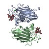
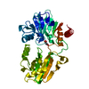
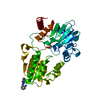
Mass: 19589.703 Da / Num. of mol.: 2 / Fragment: RESIDUES 37-190 Source method: isolated from a genetically manipulated source Source: (gene. exp.) VACCINIA VIRUS / Strain: WESTERN RESERVE / Plasmid: POPINF / Production host: ESCHERICHIA COLI (E. coli) / Strain (production host): ROSETTA(DE3) PLYSS / References: UniProt: Q01220
Sequence details
RESIDUES 1-36 ARE REMOVED, AN N-TERMINAL START CODON IS ADDED AND A C-TERMINAL LYS-HIS6 ...RESIDUES 1-36 ARE REMOVED, AN N-TERMINAL START CODON IS ADDED AND A C-TERMINAL LYS-HIS6 PURIFICATION TAG IS ADDED.
-
Experimental details
-
Experiment
Experiment
Method: X-RAY DIFFRACTION / Number of used crystals: 1
-
Sample preparation
Crystal
Density Matthews: 4.88 Å3/Da / Density % sol: 74.83 % Description: MODEL WAS STRIPPED OF SOLVENT AND ALTERNATE CONFORMATIONS PRIOR TO MOLECULAR REPLACEMENT.
Crystal grow
Temperature: 294 K / Method: vapor diffusion, hanging drop Details: HANGING DROPS CONTAINING 1 UL PROTEIN (5.4 MG/ML IN 20 MM TRIS, 200 MM NACL, PH 7.5) AND 1 UL RESERVOIR SOLUTION (0.2 M SODIUM PHOSPHATE, 22% (W/V) PEG 3350) WERE EQUILIBRATED AGAINST 500 UL ...Details: HANGING DROPS CONTAINING 1 UL PROTEIN (5.4 MG/ML IN 20 MM TRIS, 200 MM NACL, PH 7.5) AND 1 UL RESERVOIR SOLUTION (0.2 M SODIUM PHOSPHATE, 22% (W/V) PEG 3350) WERE EQUILIBRATED AGAINST 500 UL RESERVOIRS AT 21 C. CRYSTALS WERE CRYOPROTECTED IN RESERVOIR SOLUTION SUPPLEMENTED WITH 20% (V/V) GLYCEROL.
In the structure databanks used in Yorodumi, some data are registered as the other names, "COVID-19 virus" and "2019-nCoV". Here are the details of the virus and the list of structure data.
Jan 31, 2019. EMDB accession codes are about to change! (news from PDBe EMDB page)
EMDB accession codes are about to change! (news from PDBe EMDB page)
The allocation of 4 digits for EMDB accession codes will soon come to an end. Whilst these codes will remain in use, new EMDB accession codes will include an additional digit and will expand incrementally as the available range of codes is exhausted. The current 4-digit format prefixed with “EMD-” (i.e. EMD-XXXX) will advance to a 5-digit format (i.e. EMD-XXXXX), and so on. It is currently estimated that the 4-digit codes will be depleted around Spring 2019, at which point the 5-digit format will come into force.
The EM Navigator/Yorodumi systems omit the EMD- prefix.
Related info.:Q: What is EMD? / ID/Accession-code notation in Yorodumi/EM Navigator
Yorodumi is a browser for structure data from EMDB, PDB, SASBDB, etc.
This page is also the successor to EM Navigator detail page, and also detail information page/front-end page for Omokage search.
The word "yorodu" (or yorozu) is an old Japanese word meaning "ten thousand". "mi" (miru) is to see.
Related info.:EMDB / PDB / SASBDB / Comparison of 3 databanks / Yorodumi Search / Aug 31, 2016. New EM Navigator & Yorodumi / Yorodumi Papers / Jmol/JSmol / Function and homology information / Changes in new EM Navigator and Yorodumi
 Movie
Movie Controller


 Open data
 Basic information
 Components
 Keywords
 Function and homology information
 VACCINIA VIRUS
 X-RAY DIFFRACTION /
  Authors
 Citation
 Structure visualization
 Downloads & links
 Other downloads


 PDBj
PDBj Assembly
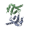


 Sample preparation
 / Beamline: I03 / Wavelength: 1.06
  Processing