[English] 日本語
Yorodumi
- PDB-2vvy: Structure of Vaccinia virus protein B14 -

+
Open data


ID or keywords:

Loading...

-
Basic information

Entry
Database: PDB / ID: 2vvy
TitleStructure of Vaccinia virus protein B14
ComponentsPROTEIN B15
KeywordsVIRAL PROTEIN / IKK / IKK BETA / BCL-2 FAMILY / EARLY PROTEIN / HOST-VIRUS INTERACTION / VACCINIA VIRUS / IMMUNOMODULATOR / NF-KB ACTIVATION
Function / homology
Function and homology information


Poxvirus, Protein B14/B22/C16 / dsDNA poxvirus / Poxvirus Bcl-2-like / Poxvirus domain superfamily / Poxvirus Bcl-2-like proteins / Apoptosis Regulator Bcl-x / Orthogonal Bundle / Mainly Alpha
Similarity search - Domain/homology
Biological speciesVACCINIA VIRUS
MethodX-RAY DIFFRACTION / SYNCHROTRON / MAD / Resolution: 2.693 Å
AuthorsGraham, S.C. / Bahar, M.W. / Cooray, S. / Chen, R.A.-J. / Whalen, D.M. / Abrescia, N.G.A. / Alderton, D. / Owens, R.J. / Stuart, D.I. / Smith, G.L. / Grimes, J.M.
CitationJournal: Plos Pathog. / Year: 2008
Title: Vaccinia Virus Proteins A52 and B14 Share a Bcl-2-Like Fold But Have Evolved to Inhibit NF-kappaB Rather Than Apoptosis
Authors: Graham, S.C. / Bahar, M.W. / Cooray, S. / Chen, R.A.-J. / Whalen, D.M. / Abrescia, N.G.A. / Alderton, D. / Owens, R.J. / Stuart, D.I. / Smith, G.L. / Grimes, J.M.
History
DepositionJun 12, 2008Deposition site: PDBE / Processing site: PDBE
Revision 1.0Aug 26, 2008Provider: repository / Type: Initial release
Revision 1.1May 8, 2011Group: Version format compliance
Revision 1.2Jul 13, 2011Group: Version format compliance
Revision 1.3May 8, 2019Group: Data collection / Derived calculations ...Data collection / Derived calculations / Experimental preparation / Other
Category: database_PDB_rev / database_PDB_rev_record ...database_PDB_rev / database_PDB_rev_record / exptl_crystal_grow / pdbx_database_proc / pdbx_database_status / struct_conn
Item: _exptl_crystal_grow.method / _exptl_crystal_grow.temp ..._exptl_crystal_grow.method / _exptl_crystal_grow.temp / _pdbx_database_status.recvd_author_approval / _struct_conn.pdbx_leaving_atom_flag
Remark 650 HELIX DETERMINATION METHOD: AUTHOR PROVIDED.

-
Structure visualization

Structure viewerMolecule:
MolmilJmol/JSmol

Downloads & links

-
Assembly

Deposited unit
A: PROTEIN B15
B: PROTEIN B15
C: PROTEIN B15
D: PROTEIN B15


Theoretical massNumber of molelcules
Total (without water)79,4024
Polymers79,4024
Non-polymers00
Water00
1
A: PROTEIN B15
C: PROTEIN B15


Theoretical massNumber of molelcules
Total (without water)39,7012
Polymers39,7012
Non-polymers00
Water0
TypeNameSymmetry operationNumber
identity operation1_555x,y,z1
Buried area2020 Å2
ΔGint-13.7 kcal/mol
Surface area15690 Å2
MethodPQS
2
B: PROTEIN B15
D: PROTEIN B15


Theoretical massNumber of molelcules
Total (without water)39,7012
Polymers39,7012
Non-polymers00
Water0
TypeNameSymmetry operationNumber
identity operation1_555x,y,z1
Buried area1810 Å2
ΔGint-10.9 kcal/mol
Surface area15560 Å2
MethodPQS
Unit cell
Length a, b, c (Å)168.673, 43.884, 99.878
Angle α, β, γ (deg.)90.00, 112.09, 90.00
Int Tables number5
Space group name H-MC121
Noncrystallographic symmetry (NCS)NCS domain:
IDEns-ID
11
21
31
41

NCS domain segments:
Dom-IDComponent-IDEns-IDSelection details
111CHAIN A
211CHAIN B AND NOT (RESID 1:9 OR RESID 27:28 OR RESID 42:45 OR RESID 117:120 )
311CHAIN C AND NOT (RESID 1:9 OR RESID 27:28 OR RESID 42:45 OR RESID 117:120 )
411CHAIN D AND NOT (RESID 1:9 OR RESID 27:28 OR RESID 42:45 OR RESID 117:120 )

-
Components

#1: Protein
PROTEIN B15 / B14


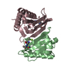
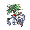
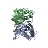
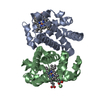
Mass: 19850.463 Da / Num. of mol.: 4
Source method: isolated from a genetically manipulated source
Source: (gene. exp.) VACCINIA VIRUS / Strain: WESTERN RESERVE / Plasmid: PET-28A / Production host: ESCHERICHIA COLI (E. coli) / Strain (production host): B834(DE3) / References: UniProt: P24772
Sequence detailsTHE ADDED N-TERMINAL HIS6 FUSION TAG AND THROMBIN CLEAVAGE SITE ARISE FROM THE PET-28A EXPRESSION VECTOR.

-
Experimental details

-
Experiment

ExperimentMethod: X-RAY DIFFRACTION / Number of used crystals: 1

-
Sample preparation

CrystalDensity Matthews: 2.2 Å3/Da / Density % sol: 44.1 % / Description: NONE
Crystal growTemperature: 294 K / Method: vapor diffusion, sitting drop
Details: SITTING DROPS CONTAINING 100 NL PROTEIN (3.2 MG/ML IN 50 MM TRIS PH 8.5, 150 MM NACL, 2 MM 2-MERCAPTOETHANOL) AND 100 NL RESERVOIR SOLUTION (0.2 M DIAMMONIUM TARTRATE, 20% (W/V) PEG 3350, 0. ...Details: SITTING DROPS CONTAINING 100 NL PROTEIN (3.2 MG/ML IN 50 MM TRIS PH 8.5, 150 MM NACL, 2 MM 2-MERCAPTOETHANOL) AND 100 NL RESERVOIR SOLUTION (0.2 M DIAMMONIUM TARTRATE, 20% (W/V) PEG 3350, 0.4 M NON-DETERGENT SULPHOBETANE 201 AND 2 MM 2-MERCAPTOETHANOL) WERE EQUILIBRATED AGAINST 95 UL RESERVOIRS AT 21 C. CRYSTALS WERE CRYOPROTECTED IN RESERVOIR SOLUTION SUPPLEMENTED WITH 20% (V/V) GLYCEROL.

-
Data collection

DiffractionMean temperature: 100 K
Diffraction sourceSource: SYNCHROTRON / Site: ESRF / Beamline: BM14 / Wavelength: 0.8856
DetectorType: MARRESEARCH / Detector: CCD / Date: Jul 8, 2005 / Details: MIRRORS
RadiationMonochromator: SI(111) CRYSTAL / Protocol: SINGLE WAVELENGTH / Monochromatic (M) / Laue (L): M / Scattering type: x-ray
Radiation wavelengthWavelength: 0.8856 Å / Relative weight: 1
ReflectionResolution: 2.7→50 Å / Num. obs: 19127 / % possible obs: 99.8 % / Observed criterion σ(I): -3 / Redundancy: 3.7 % / Biso Wilson estimate: 50.56 Å2 / Rmerge(I) obs: 0.12 / Net I/σ(I): 10.4
Reflection shellResolution: 2.7→2.8 Å / Redundancy: 3.4 % / Rmerge(I) obs: 0.84 / Mean I/σ(I) obs: 1.8 / % possible all: 98.6

-
Processing

Software
NameVersionClassification
PHENIX(PHENIX.REFINE)refinement
HKL-2000data reduction
HKL-2000data scaling
HKL2MAPphasing
RefinementMethod to determine structure: MAD
Starting model: NONE

Resolution: 2.693→46.273 Å / SU ML: 0.35 / Phase error: 28.66 / Stereochemistry target values: ML / Details: B VALUES INCLUDE TLS CONTRIBUTIONS.
RfactorNum. reflection% reflection
Rfree0.2439 1783 4.9 %
Rwork0.2225 --
obs0.2236 36439 99.23 %
Solvent computationShrinkage radii: 0.9 Å / VDW probe radii: 1.11 Å / Solvent model: FLAT BULK SOLVENT MODEL / Bsol: 39.43 Å2 / ksol: 0.365 e/Å3
Displacement parametersBiso mean: 62.5 Å2
Baniso -1Baniso -2Baniso -3
1--10.5475 Å20 Å22.6113 Å2
2---8.2398 Å20 Å2
3---13.8357 Å2
Refinement stepCycle: LAST / Resolution: 2.693→46.273 Å
ProteinNucleic acidLigandSolventTotal
Num. atoms4632 0 0 0 4632
Refine LS restraints
Refine-IDTypeDev idealNumber
X-RAY DIFFRACTIONf_bond_d0.0054744
X-RAY DIFFRACTIONf_angle_d0.7776440
X-RAY DIFFRACTIONf_dihedral_angle_d14.1631680
X-RAY DIFFRACTIONf_chiral_restr0.054700
X-RAY DIFFRACTIONf_plane_restr0.004828
Refine LS restraints NCS
Ens-IDDom-IDAuth asym-IDNumberRefine-IDTypeRms dev position (Å)
11A1051X-RAY DIFFRACTIONPOSITIONAL
12B1051X-RAY DIFFRACTIONPOSITIONAL0.029
13C1051X-RAY DIFFRACTIONPOSITIONAL0.026
14D1047X-RAY DIFFRACTIONPOSITIONAL0.028
LS refinement shell
Resolution (Å)Rfactor RfreeNum. reflection RfreeRfactor RworkNum. reflection RworkRefine-ID% reflection obs (%)
2.6934-2.76620.4123990.38082577X-RAY DIFFRACTION94
2.7662-2.84760.4131250.36122659X-RAY DIFFRACTION100
2.8476-2.93950.35371670.33752713X-RAY DIFFRACTION100
2.9395-3.04450.28531700.30262600X-RAY DIFFRACTION100
3.0445-3.16640.24161260.27222689X-RAY DIFFRACTION100
3.1664-3.31040.27521390.25922709X-RAY DIFFRACTION100
3.3104-3.48490.28081610.24522653X-RAY DIFFRACTION100
3.4849-3.70320.29871440.22722691X-RAY DIFFRACTION100
3.7032-3.98890.23531620.19342659X-RAY DIFFRACTION100
3.9889-4.39010.17721210.16662716X-RAY DIFFRACTION100
4.3901-5.02470.1951110.15962679X-RAY DIFFRACTION100
5.0247-6.3280.19811070.18052711X-RAY DIFFRACTION99
6.328-46.27930.16461510.17162600X-RAY DIFFRACTION98
Refinement TLS params.

Method: refined / Refine-ID: X-RAY DIFFRACTION

IDL112)L122)L132)L222)L232)L332)S11 (Å °)S12 (Å °)S13 (Å °)S21 (Å °)S22 (Å °)S23 (Å °)S31 (Å °)S32 (Å °)S33 (Å °)T112)T122)T132)T222)T232)T332)Origin x (Å)Origin y (Å)Origin z (Å)
11.095-0.1026-0.36642.0780.3120.64040.0264-0.13380.02560.0852-0.1448-0.12450.049-0.01790.08260.0422-0.050.03410.17760.00680.1204-40.08712.777612.7107
23.57251.0957-1.0984-0.3931-0.61581.6448-0.05690.0681-0.5158-0.0849-0.14090.01740.21760.03720.21280.29010.0556-0.11710.1429-0.02370.427-79.8836-2.753919.7358
33.52030.637-0.37850.0122-0.20622.1519-0.0942-0.2057-0.3994-0.13110.00330.09010.2067-0.09250.08490.24560.0315-0.06940.16630.03980.4462-20.8903-3.043525.5946
42.52580.08720.37490.8843-0.15273.2589-0.0150.055-0.0019-0.053-0.0692-0.0463-0.18960.04910.00360.20750.0349-0.03620.0886-0.01030.1211-62.014.210933.3564
Refinement TLS group
IDRefine-IDRefine TLS-IDSelection details
1X-RAY DIFFRACTION1CHAIN A
2X-RAY DIFFRACTION2CHAIN B
3X-RAY DIFFRACTION3CHAIN C
4X-RAY DIFFRACTION4CHAIN D

+
About Yorodumi

-
News

-
Feb 9, 2022. New format data for meta-information of EMDB entries

New format data for meta-information of EMDB entries

  • Version 3 of the EMDB header file is now the official format.
  • The previous official version 1.9 will be removed from the archive.

Related info.:EMDB header

External links:wwPDB to switch to version 3 of the EMDB data model

-
Aug 12, 2020. Covid-19 info

Covid-19 info

URL: https://pdbjlvh1.pdbj.org/emnavi/covid19.php

New page: Covid-19 featured information page in EM Navigator.

Related info.:Covid-19 info / Mar 5, 2020. Novel coronavirus structure data

+
Mar 5, 2020. Novel coronavirus structure data

Novel coronavirus structure data

Related info.:Yorodumi Speices / Aug 12, 2020. Covid-19 info

External links:COVID-19 featured content - PDBj / Molecule of the Month (242):Coronavirus Proteases

+
Jan 31, 2019. EMDB accession codes are about to change! (news from PDBe EMDB page)

EMDB accession codes are about to change! (news from PDBe EMDB page)

  • The allocation of 4 digits for EMDB accession codes will soon come to an end. Whilst these codes will remain in use, new EMDB accession codes will include an additional digit and will expand incrementally as the available range of codes is exhausted. The current 4-digit format prefixed with “EMD-” (i.e. EMD-XXXX) will advance to a 5-digit format (i.e. EMD-XXXXX), and so on. It is currently estimated that the 4-digit codes will be depleted around Spring 2019, at which point the 5-digit format will come into force.
  • The EM Navigator/Yorodumi systems omit the EMD- prefix.

Related info.:Q: What is EMD? / ID/Accession-code notation in Yorodumi/EM Navigator

External links:EMDB Accession Codes are Changing Soon! / Contact to PDBj

+
Jul 12, 2017. Major update of PDB

Major update of PDB

  • wwPDB released updated PDB data conforming to the new PDBx/mmCIF dictionary.
  • This is a major update changing the version number from 4 to 5, and with Remediation, in which all the entries are updated.
  • In this update, many items about electron microscopy experimental information are reorganized (e.g. em_software).
  • Now, EM Navigator and Yorodumi are based on the updated data.

External links:wwPDB Remediation / Enriched Model Files Conforming to OneDep Data Standards Now Available in the PDB FTP Archive

-
Yorodumi

Thousand views of thousand structures

  • Yorodumi is a browser for structure data from EMDB, PDB, SASBDB, etc.
  • This page is also the successor to EM Navigator detail page, and also detail information page/front-end page for Omokage search.
  • The word "yorodu" (or yorozu) is an old Japanese word meaning "ten thousand". "mi" (miru) is to see.

Related info.:EMDB / PDB / SASBDB / Comparison of 3 databanks / Yorodumi Search / Aug 31, 2016. New EM Navigator & Yorodumi / Yorodumi Papers / Jmol/JSmol / Function and homology information / Changes in new EM Navigator and Yorodumi

Read more