 Movie
Movie Controller


+ Open data

- Basic information
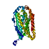
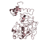
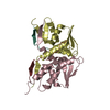
| Entry | Database: PDB / ID: 1pci | ||||||
|---|---|---|---|---|---|---|---|
| Title | PROCARICAIN | ||||||
 Components Components | PROCARICAIN | ||||||
 Keywords Keywords | THIOL PROTEASE / ZYMOGEN / HYDROLASE | ||||||
| Function / homology |  Function and homology information Function and homology information | ||||||
| Biological species |  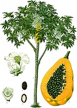 Carica papaya (papaya) Carica papaya (papaya) | ||||||
| Method |  X-RAY DIFFRACTION / SYNCHROTRON / COMBINED MOLECULAR REPLACEMENT, ISOMORPHOUS REPLACEMENT / Resolution: 3.2 Å X-RAY DIFFRACTION / SYNCHROTRON / COMBINED MOLECULAR REPLACEMENT, ISOMORPHOUS REPLACEMENT / Resolution: 3.2 Å | ||||||
 Authors Authors | Groves, M.R. / Taylor, M.A.J. / Scott, M. / Cummings, N.J. / Pickersgill, R.W. / Jenkins, J.A. | ||||||
 Citation Citation | Journal: Structure / Year: 1996 Title: The prosequence of procaricain forms an alpha-helical domain that prevents access to the substrate-binding cleft. Authors: Groves, M.R. / Taylor, M.A. / Scott, M. / Cummings, N.J. / Pickersgill, R.W. / Jenkins, J.A. #1: Journal: Acta Crystallogr.,Sect.B / Year: 1991Title: Determination of the Structure of Papaya Protease Omega Authors: Pickersgill, R.W. / Rizkallah, P. / Harris, G.W. / Goodenough, P.W. | ||||||
| History |
|
- Structure visualization
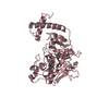
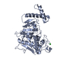
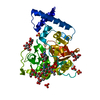
| Structure viewer | Molecule: MolmilJmol/JSmol |
|---|
- Downloads & links
-Download
| PDBx/mmCIF format | 1pci.cif.gz | 172.6 KB | Display | PDBx/mmCIF format |
|---|---|---|---|---|
| PDB format | pdb1pci.ent.gz | 141.2 KB | Display | PDB format |
| PDBx/mmJSON format | 1pci.json.gz | Tree view | PDBx/mmJSON format | |
| Others |  Other downloads Other downloads |
-Validation report
| Arichive directory | https://data.pdbj.org/pub/pdb/validation_reports/pc/1pciftp://data.pdbj.org/pub/pdb/validation_reports/pc/1pci | HTTPS FTP |
|---|
-Related structure data
| Related structure data |  1ppoS S: Starting model for refinement |
|---|---|
| Similar structure data |


-Links
 PDBj
PDBj
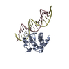
- Assembly
| Deposited unit | 
| ||||||||||||
|---|---|---|---|---|---|---|---|---|---|---|---|---|---|
| 1 | 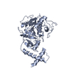
| ||||||||||||
| 2 | 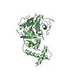
| ||||||||||||
| 3 | 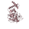
| ||||||||||||
| Unit cell |
| ||||||||||||
| Noncrystallographic symmetry (NCS) | NCS oper:
| ||||||||||||
| Details | THE DEPOSITOR PROVIDED COORDINATES FOR CHAIN A. THE OTHER TWO CHAINS IN THE ASYMMETRIC UNIT WERE GENERATED BY THE PROTEIN DATA BANK USING THE MTRIX TRANSFORMATIONS BELOW. |
-Components
| #1: Protein | Mass: 35940.328 Da / Num. of mol.: 3 / Mutation: H159G Source method: isolated from a genetically manipulated source Details: ACTIVE SITE HIS MUTATION / Source: (gene. exp.) Carica papaya (papaya) / Tissue: LEAF / Gene: OMEGA / Plasmid: PET3A / Gene (production host): OMEGA / Production host:  Has protein modification | Y | Source details | PROCARICAI | |
|---|
-Experimental details
-Experiment
| Experiment | Method: X-RAY DIFFRACTION / Number of used crystals: 5 |
|---|
- Sample preparation
| Crystal | Density Matthews: 3.84 Å3/Da / Density % sol: 62 % | ||||||||||||||||||||||||||||||||||||||||
|---|---|---|---|---|---|---|---|---|---|---|---|---|---|---|---|---|---|---|---|---|---|---|---|---|---|---|---|---|---|---|---|---|---|---|---|---|---|---|---|---|---|
| Crystal grow | pH: 7 / Details: pH 7. | ||||||||||||||||||||||||||||||||||||||||
| Crystal grow | *PLUS Temperature: 11 ℃ / pH: 5 / Method: vapor diffusion, hanging drop | ||||||||||||||||||||||||||||||||||||||||
| Components of the solutions | *PLUS
|
-Data collection
| Diffraction | Mean temperature: 100 K | |||||||||
|---|---|---|---|---|---|---|---|---|---|---|
| Diffraction source | Source: SYNCHROTRON / Site: SRS  / Beamline: PX9.6 / Wavelength: 0.87, 1.0 / Beamline: PX9.6 / Wavelength: 0.87, 1.0 | |||||||||
| Detector | Type: MARRESEARCH / Detector: IMAGE PLATE / Date: Dec 23, 1994 | |||||||||
| Radiation | Monochromatic (M) / Laue (L): M / Scattering type: x-ray | |||||||||
| Radiation wavelength |
| |||||||||
| Reflection | Resolution: 3.2→96.2 Å / Num. obs: 27079 / % possible obs: 98.6 % / Observed criterion σ(I): 1 / Redundancy: 5.1 % / Biso Wilson estimate: 0.29 Å2 / Rmerge(I) obs: 0.086 / Net I/σ(I): 3.8 | |||||||||
| Reflection shell | Resolution: 3.21→3.34 Å / Redundancy: 5.1 % / Rmerge(I) obs: 0.169 / % possible all: 98.6 | |||||||||
| Reflection shell | *PLUS % possible obs: 98.6 % |
- Processing
| Software |
| ||||||||||||||||||||||
|---|---|---|---|---|---|---|---|---|---|---|---|---|---|---|---|---|---|---|---|---|---|---|---|
| Refinement | Method to determine structure: COMBINED MOLECULAR REPLACEMENT, ISOMORPHOUS REPLACEMENT Starting model: MATURE CARICAIN (PDB ENTRY 1PPO) Resolution: 3.2→90.6 Å / Rfactor Rfree error: 0.0075 / Cross valid method: FREE R / σ(F): 1 Details: ELECTRON DENSITY FOR THE POLYPEPTIDE CHAIN 90P - 103P IS POOR. THE STRUCTURE WAS PRIMARILY MODELED IN THIS REGION. NCS RESTRAINTS WERE RELAXED FOR THE POLYPEPTIDE CHAIN 88P - 100P DUE TO NCS PACKING CLASHES.
| ||||||||||||||||||||||
| Displacement parameters | Biso mean: 0.94 Å2
| ||||||||||||||||||||||
| Refine analyze | Luzzati d res low obs: 96.2 Å | ||||||||||||||||||||||
| Refinement step | Cycle: LAST / Resolution: 3.2→90.6 Å
| ||||||||||||||||||||||
| Refine LS restraints NCS | Weight Biso : 1 / Weight position: 300 | ||||||||||||||||||||||
| LS refinement shell | Resolution: 3.2→3.22 Å / Rfactor Rfree error: 0.0072
| ||||||||||||||||||||||
| Xplor file | Serial no: 1 / Param file: PARHCSDX.PRO / Topol file: TPOH19.PEP | ||||||||||||||||||||||
| Software | *PLUS Name: X-PLOR / Classification: refinement | ||||||||||||||||||||||
| Refinement | *PLUS | ||||||||||||||||||||||
| Solvent computation | *PLUS | ||||||||||||||||||||||
| Displacement parameters | *PLUS Biso mean: 53.7 Å2 |
