 Movie
Movie Controller


+ Open data

- Basic information
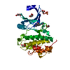
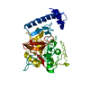
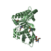
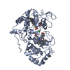
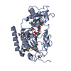
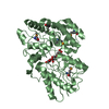
| Entry | Database: PDB / ID: 6czs | |||||||||
|---|---|---|---|---|---|---|---|---|---|---|
| Title | Crystal structure of human pro-cathepsin H C26S mutant | |||||||||
 Components Components | Pro-cathepsin H | |||||||||
 Keywords Keywords | HYDROLASE / papain family cysteine peptidase / protein degradation in lysosome / inhibitory prodomain | |||||||||
| Function / homology |  Function and homology information Function and homology informationcathepsin H / dichotomous subdivision of terminal units involved in lung branching / HLA-A specific activating MHC class I receptor activity / kininogen binding / neuropeptide catabolic process / multivesicular body lumen / outer dense fiber / alveolar lamellar body / immune response-regulating signaling pathway / response to odorant ...cathepsin H / dichotomous subdivision of terminal units involved in lung branching / HLA-A specific activating MHC class I receptor activity / kininogen binding / neuropeptide catabolic process / multivesicular body lumen / outer dense fiber / alveolar lamellar body / immune response-regulating signaling pathway / response to odorant / thyroid hormone binding / membrane protein proteolysis / lysosomal protein catabolic process / bradykinin catabolic process / metanephros development / surfactant homeostasis / Surfactant metabolism / cellular response to thyroid hormone stimulus / zymogen activation / antigen processing and presentation / cysteine-type endopeptidase activator activity involved in apoptotic process / positive regulation of epithelial cell migration / response to retinoic acid / aminopeptidase activity / axoneme / ERK1 and ERK2 cascade / cysteine-type peptidase activity / MHC class II antigen presentation / acrosomal vesicle / : / positive regulation of apoptotic signaling pathway / T cell mediated cytotoxicity / protein destabilization / tertiary granule lumen / peptidase activity / extracellular matrix / secretory granule lumen / endopeptidase activity / spermatogenesis / ficolin-1-rich granule lumen / adaptive immune response / lysosome / immune response / positive regulation of cell migration / serine-type endopeptidase activity / cysteine-type endopeptidase activity / positive regulation of gene expression / Neutrophil degranulation / protein-containing complex binding / proteolysis / : / extracellular exosome / extracellular region / identical protein binding / cytosol Similarity search - Function | |||||||||
| Biological species |  Homo sapiens (human) Homo sapiens (human) | |||||||||
| Method |  X-RAY DIFFRACTION / SYNCHROTRON / MOLECULAR REPLACEMENT / Resolution: 1.66 Å X-RAY DIFFRACTION / SYNCHROTRON / MOLECULAR REPLACEMENT / Resolution: 1.66 Å | |||||||||
 Authors Authors | Huang, X. / Hao, Y. | |||||||||
 Citation Citation | Journal: PLoS ONE / Year: 2018 Title: Crystal structures of human procathepsin H. Authors: Hao, Y. / Purtha, W. / Cortesio, C. / Rui, H. / Gu, Y. / Chen, H. / Sickmier, E.A. / Manzanillo, P. / Huang, X. | |||||||||
| History |
|
- Structure visualization
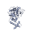
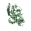
| Structure viewer | Molecule: MolmilJmol/JSmol |
|---|
- Downloads & links
-Download
| PDBx/mmCIF format | 6czs.cif.gz | 95.9 KB | Display | PDBx/mmCIF format |
|---|---|---|---|---|
| PDB format | pdb6czs.ent.gz | 70.1 KB | Display | PDB format |
| PDBx/mmJSON format | 6czs.json.gz | Tree view | PDBx/mmJSON format | |
| Others |  Other downloads Other downloads |
-Validation report
| Arichive directory | https://data.pdbj.org/pub/pdb/validation_reports/cz/6czsftp://data.pdbj.org/pub/pdb/validation_reports/cz/6czs | HTTPS FTP |
|---|
-Related structure data
| Related structure data |  6czkC  8pchS S: Starting model for refinement C: citing same article ( |
|---|---|
| Similar structure data |


-Links
 PDBj
PDBj


- Assembly
| Deposited unit | 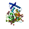
| ||||||||||||
|---|---|---|---|---|---|---|---|---|---|---|---|---|---|
| 1 |
| ||||||||||||
| Unit cell |
| ||||||||||||
| Components on special symmetry positions |
|
-Components
-Protein , 1 types, 1 molecules A
| #1: Protein | Mass: 37417.492 Da / Num. of mol.: 1 / Mutation: C26S Source method: isolated from a genetically manipulated source Source: (gene. exp.) Homo sapiens (human) / Gene: CTSH, CPSB / Production host:   Spodoptera frugiperda (fall armyworm) / References: UniProt: P09668, cathepsin H Spodoptera frugiperda (fall armyworm) / References: UniProt: P09668, cathepsin H |
|---|
-Sugars , 3 types, 4 molecules 
| #2: Polysaccharide | alpha-D-mannopyranose-(1-3)-[alpha-D-mannopyranose-(1-6)]beta-D-mannopyranose-(1-4)-2-acetamido-2- ...alpha-D-mannopyranose-(1-3)-[alpha-D-mannopyranose-(1-6)]beta-D-mannopyranose-(1-4)-2-acetamido-2-deoxy-beta-D-glucopyranose-(1-4)-2-acetamido-2-deoxy-beta-D-glucopyranose Source method: isolated from a genetically manipulated source | ||
|---|---|---|---|
| #3: Polysaccharide | 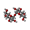  Source method: isolated from a genetically manipulated source Details: oligosaccharide with reducing-end-to-reducing-end glycosidic bond References: trehalose #4: Sugar | ChemComp-NAG / |  Type: D-saccharide, beta linking / Mass: 221.208 Da / Num. of mol.: 1 Type: D-saccharide, beta linking / Mass: 221.208 Da / Num. of mol.: 1Source method: isolated from a genetically manipulated source Formula: C8H15NO6 |
-Non-polymers , 3 types, 505 molecules 


| #5: Chemical | ChemComp-SO4 /  Mass: 96.063 Da / Num. of mol.: 5 / Source method: obtained synthetically / Formula: SO4 Mass: 96.063 Da / Num. of mol.: 5 / Source method: obtained synthetically / Formula: SO4#6: Chemical |  Mass: 35.453 Da / Num. of mol.: 2 / Source method: obtained synthetically / Formula: Cl Mass: 35.453 Da / Num. of mol.: 2 / Source method: obtained synthetically / Formula: Cl#7: Water | ChemComp-HOH / | Mass: 18.015 Da / Num. of mol.: 498 / Source method: isolated from a natural source / Formula: H2O |
|---|
-Details
| Has protein modification | Y |
|---|
-Experimental details
-Experiment
| Experiment | Method: X-RAY DIFFRACTION / Number of used crystals: 1 |
|---|
- Sample preparation
| Crystal | Density Matthews: 3.97 Å3/Da / Density % sol: 69 % |
|---|---|
| Crystal grow | Temperature: 293 K / Method: vapor diffusion, sitting drop / pH: 4.2 Details: 0.1 M sodium phosphate dibasic / citric acid, pH 4.2, 2.0 M ammonium sulfate |
-Data collection
| Diffraction | Mean temperature: 100 K |
|---|---|
| Diffraction source | Source: SYNCHROTRON / Site: ALS  / Beamline: 5.0.2 / Wavelength: 1 Å / Beamline: 5.0.2 / Wavelength: 1 Å |
| Detector | Type: DECTRIS PILATUS3 6M / Detector: PIXEL / Date: Jun 16, 2017 |
| Radiation | Protocol: SINGLE WAVELENGTH / Monochromatic (M) / Laue (L): M / Scattering type: x-ray |
| Radiation wavelength | Wavelength: 1 Å / Relative weight: 1 |
| Reflection | Resolution: 1.66→49.12 Å / Num. obs: 70589 / % possible obs: 100 % / Redundancy: 19.3 % / Rmerge(I) obs: 0.087 / Rpim(I) all: 0.02 / Rrim(I) all: 0.089 / Net I/σ(I): 20.4 |
| Reflection shell | Resolution: 1.66→1.69 Å / Redundancy: 20.4 % / Rmerge(I) obs: 1.658 / Mean I/σ(I) obs: 2.2 / Num. unique obs: 3478 / Rpim(I) all: 0.376 / Rrim(I) all: 1.7 / % possible all: 100 |
- Processing
| Software |
| |||||||||||||||||||||||||||||||||||||||||||||||||||||||||||||||||||||||||||||||||||||||||||||||||||||||||||||||||||||||||||||||||||||||||||||||||||||||||||||||||||||||||||||||
|---|---|---|---|---|---|---|---|---|---|---|---|---|---|---|---|---|---|---|---|---|---|---|---|---|---|---|---|---|---|---|---|---|---|---|---|---|---|---|---|---|---|---|---|---|---|---|---|---|---|---|---|---|---|---|---|---|---|---|---|---|---|---|---|---|---|---|---|---|---|---|---|---|---|---|---|---|---|---|---|---|---|---|---|---|---|---|---|---|---|---|---|---|---|---|---|---|---|---|---|---|---|---|---|---|---|---|---|---|---|---|---|---|---|---|---|---|---|---|---|---|---|---|---|---|---|---|---|---|---|---|---|---|---|---|---|---|---|---|---|---|---|---|---|---|---|---|---|---|---|---|---|---|---|---|---|---|---|---|---|---|---|---|---|---|---|---|---|---|---|---|---|---|---|---|---|---|
| Refinement | Method to determine structure: MOLECULAR REPLACEMENT Starting model: 8PCH Resolution: 1.66→49.121 Å / SU ML: 0.2 / Cross valid method: FREE R-VALUE / σ(F): 1.35 / Phase error: 19.79 / Stereochemistry target values: ML
| |||||||||||||||||||||||||||||||||||||||||||||||||||||||||||||||||||||||||||||||||||||||||||||||||||||||||||||||||||||||||||||||||||||||||||||||||||||||||||||||||||||||||||||||
| Solvent computation | Shrinkage radii: 0.9 Å / VDW probe radii: 1.11 Å / Solvent model: FLAT BULK SOLVENT MODEL | |||||||||||||||||||||||||||||||||||||||||||||||||||||||||||||||||||||||||||||||||||||||||||||||||||||||||||||||||||||||||||||||||||||||||||||||||||||||||||||||||||||||||||||||
| Refinement step | Cycle: LAST / Resolution: 1.66→49.121 Å
| |||||||||||||||||||||||||||||||||||||||||||||||||||||||||||||||||||||||||||||||||||||||||||||||||||||||||||||||||||||||||||||||||||||||||||||||||||||||||||||||||||||||||||||||
| Refine LS restraints |
| |||||||||||||||||||||||||||||||||||||||||||||||||||||||||||||||||||||||||||||||||||||||||||||||||||||||||||||||||||||||||||||||||||||||||||||||||||||||||||||||||||||||||||||||
| LS refinement shell |
|
