 Movie
Movie Controller

[English] 日本語
 Yorodumi
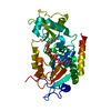
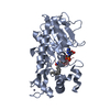
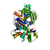
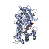
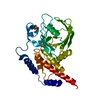
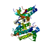
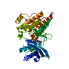
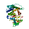
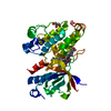
- PDB-5mar: Structure of human SIRT2 in complex with 1,2,4-Oxadiazole inhibit... -
+ Open data

- Basic information
| Entry | Database: PDB / ID: 5mar | ||||||
|---|---|---|---|---|---|---|---|
| Title | Structure of human SIRT2 in complex with 1,2,4-Oxadiazole inhibitor and ADP ribose. | ||||||
 Components Components | NAD-dependent protein deacetylase sirtuin-2 | ||||||
 Keywords Keywords | HYDROLASE / Sirtuin / NAD-dependent Protein deacylase / Inhibitor Complex | ||||||
| Function / homology |  Function and homology information Function and homology informationcellular response to caloric restriction / negative regulation of oligodendrocyte progenitor proliferation / histone methacryllysine demethacrylase activity / histone benzoyllysine debenzoylase activity / negative regulation of striated muscle tissue development / negative regulation of satellite cell differentiation / histone H4K16 deacetylase activity, NAD-dependent / positive regulation of attachment of spindle microtubules to kinetochore / peptidyl-lysine deacetylation / NAD-dependent protein demyristoylase activity ...cellular response to caloric restriction / negative regulation of oligodendrocyte progenitor proliferation / histone methacryllysine demethacrylase activity / histone benzoyllysine debenzoylase activity / negative regulation of striated muscle tissue development / negative regulation of satellite cell differentiation / histone H4K16 deacetylase activity, NAD-dependent / positive regulation of attachment of spindle microtubules to kinetochore / peptidyl-lysine deacetylation / NAD-dependent protein demyristoylase activity / NAD-dependent protein depalmitoylase activity / positive regulation of meiotic nuclear division / paranodal junction / tubulin deacetylation / lateral loop / mitotic nuclear membrane reassembly / regulation of phosphorylation / tubulin deacetylase activity / NLRP3 inflammasome complex assembly / negative regulation of NLRP3 inflammasome complex assembly / paranode region of axon / regulation of exit from mitosis / negative regulation of peptidyl-threonine phosphorylation / Schmidt-Lanterman incisure / positive regulation of fatty acid biosynthetic process / protein acetyllysine N-acetyltransferase / protein deacetylation / NAD-dependent protein lysine deacetylase activity / histone deacetylase activity, NAD-dependent / myelination in peripheral nervous system / rDNA heterochromatin formation / positive regulation of oocyte maturation / juxtaparanode region of axon / Initiation of Nuclear Envelope (NE) Reformation / chromatin silencing complex / meiotic spindle / histone deacetylase activity / protein lysine deacetylase activity / positive regulation of DNA binding / response to redox state / regulation of myelination / histone acetyltransferase binding / negative regulation of fat cell differentiation / negative regulation of reactive oxygen species metabolic process / positive regulation of cell division / NAD+ poly-ADP-ribosyltransferase activity / NAD+ binding / positive regulation of execution phase of apoptosis / glial cell projection / subtelomeric heterochromatin formation / lipid catabolic process / heterochromatin / cellular response to epinephrine stimulus / Transferases; Acyltransferases; Transferring groups other than aminoacyl groups / substantia nigra development / epigenetic regulation of gene expression / negative regulation of autophagy / meiotic cell cycle / ubiquitin binding / centriole / negative regulation of protein catabolic process / autophagy / spindle / histone deacetylase binding / mitotic spindle / myelin sheath / positive regulation of proteasomal ubiquitin-dependent protein catabolic process / heterochromatin formation / chromosome / growth cone / cellular response to oxidative stress / midbody / cellular response to hypoxia / DNA-binding transcription factor binding / microtubule / perikaryon / proteasome-mediated ubiquitin-dependent protein catabolic process / chromosome, telomeric region / regulation of cell cycle / innate immune response / cell division / negative regulation of DNA-templated transcription / chromatin binding / centrosome / perinuclear region of cytoplasm / nucleolus / negative regulation of transcription by RNA polymerase II / positive regulation of transcription by RNA polymerase II / mitochondrion / zinc ion binding / nucleus / plasma membrane / cytoplasm / cytosol Similarity search - Function | ||||||
| Biological species |  Homo sapiens (human) Homo sapiens (human) | ||||||
| Method |  X-RAY DIFFRACTION / SYNCHROTRON / MOLECULAR REPLACEMENT / Resolution: 1.89 Å X-RAY DIFFRACTION / SYNCHROTRON / MOLECULAR REPLACEMENT / Resolution: 1.89 Å | ||||||
 Authors Authors | Moniot, S. / Steegborn, C. | ||||||
| Funding support |  Germany, 1items Germany, 1items
| ||||||
 Citation Citation | Journal: J. Med. Chem. / Year: 2017 Title: Development of 1,2,4-Oxadiazoles as Potent and Selective Inhibitors of the Human Deacetylase Sirtuin 2: Structure-Activity Relationship, X-ray Crystal Structure, and Anticancer Activity. Authors: Moniot, S. / Forgione, M. / Lucidi, A. / Hailu, G.S. / Nebbioso, A. / Carafa, V. / Baratta, F. / Altucci, L. / Giacche, N. / Passeri, D. / Pellicciari, R. / Mai, A. / Steegborn, C. / Rotili, D. | ||||||
| History |
|
- Structure visualization
| Structure viewer | Molecule: MolmilJmol/JSmol |
|---|
- Downloads & links
-Download
| PDBx/mmCIF format | 5mar.cif.gz | 382 KB | Display | PDBx/mmCIF format |
|---|---|---|---|---|
| PDB format | pdb5mar.ent.gz | 313.7 KB | Display | PDB format |
| PDBx/mmJSON format | 5mar.json.gz | Tree view | PDBx/mmJSON format | |
| Others |  Other downloads Other downloads |
-Validation report
| Arichive directory | https://data.pdbj.org/pub/pdb/validation_reports/ma/5marftp://data.pdbj.org/pub/pdb/validation_reports/ma/5mar | HTTPS FTP |
|---|
-Related structure data
| Related structure data |  3zgvS S: Starting model for refinement |
|---|---|
| Similar structure data |

-Links
 PDBj
PDBj


- Assembly
| Deposited unit | 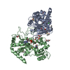
| ||||||||||||
|---|---|---|---|---|---|---|---|---|---|---|---|---|---|
| 1 | 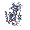
| ||||||||||||
| 2 | 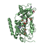
| ||||||||||||
| Unit cell |
|
-Components
-Protein , 1 types, 2 molecules AB
| #1: Protein | Mass: 34359.676 Da / Num. of mol.: 2 Source method: isolated from a genetically manipulated source Details: N-terminal residues HM are cloning artifact / Source: (gene. exp.) Homo sapiens (human) / Gene: SIRT2, SIR2L, SIR2L2 / Plasmid: pET19 modified / Details (production host): His6 SUMO N-terminal fusion tag / Production host:  References: UniProt: Q8IXJ6, Hydrolases; Acting on carbon-nitrogen bonds, other than peptide bonds; In linear amides |
|---|
-Non-polymers , 8 types, 495 molecules 
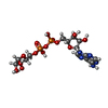
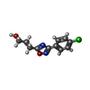


| #2: Chemical |  Mass: 65.409 Da / Num. of mol.: 2 / Source method: obtained synthetically / Formula: Zn Mass: 65.409 Da / Num. of mol.: 2 / Source method: obtained synthetically / Formula: Zn#3: Chemical |  Mass: 559.316 Da / Num. of mol.: 2 / Source method: obtained synthetically / Formula: C15H23N5O14P2 Mass: 559.316 Da / Num. of mol.: 2 / Source method: obtained synthetically / Formula: C15H23N5O14P2#4: Chemical |  Mass: 238.670 Da / Num. of mol.: 2 / Source method: obtained synthetically / Formula: C11H11ClN2O2 Mass: 238.670 Da / Num. of mol.: 2 / Source method: obtained synthetically / Formula: C11H11ClN2O2#5: Chemical |  Mass: 62.068 Da / Num. of mol.: 2 / Source method: obtained synthetically / Formula: C2H6O2 Mass: 62.068 Da / Num. of mol.: 2 / Source method: obtained synthetically / Formula: C2H6O2#6: Chemical | ChemComp-DMS /  Mass: 78.133 Da / Num. of mol.: 9 / Source method: obtained synthetically / Formula: C2H6OS / Comment: DMSO, precipitant*YM Mass: 78.133 Da / Num. of mol.: 9 / Source method: obtained synthetically / Formula: C2H6OS / Comment: DMSO, precipitant*YM#7: Chemical | ChemComp-GOL /  Mass: 92.094 Da / Num. of mol.: 4 / Source method: obtained synthetically / Formula: C3H8O3 Mass: 92.094 Da / Num. of mol.: 4 / Source method: obtained synthetically / Formula: C3H8O3#8: Chemical | ChemComp-ACT / |  Mass: 59.044 Da / Num. of mol.: 1 / Source method: obtained synthetically / Formula: C2H3O2 Mass: 59.044 Da / Num. of mol.: 1 / Source method: obtained synthetically / Formula: C2H3O2#9: Water | ChemComp-HOH / | Mass: 18.015 Da / Num. of mol.: 473 / Source method: isolated from a natural source / Formula: H2O |
|---|
-Experimental details
-Experiment
| Experiment | Method: X-RAY DIFFRACTION / Number of used crystals: 1 |
|---|
- Sample preparation
| Crystal | Density Matthews: 2.45 Å3/Da / Density % sol: 49.74 % / Description: bi-pyramidal |
|---|---|
| Crystal grow | Temperature: 293 K / Method: vapor diffusion, hanging drop / pH: 5.8 Details: 15-18 % PEG 10,000, 0.1M ammonium acetate, and 0.1M Bis-Tris pH 5.8 |
-Data collection
| Diffraction | Mean temperature: 100 K |
|---|---|
| Diffraction source | Source: SYNCHROTRON / Site: BESSY / Beamline: 14.1 / Wavelength: 0.9184 Å |
| Detector | Type: DECTRIS PILATUS 6M / Detector: PIXEL / Date: Mar 19, 2016 |
| Radiation | Monochromator: Si-111 crystal / Protocol: SINGLE WAVELENGTH / Monochromatic (M) / Laue (L): M / Scattering type: x-ray |
| Radiation wavelength | Wavelength: 0.9184 Å / Relative weight: 1 |
| Reflection | Resolution: 1.89→48.93 Å / Num. obs: 103676 / % possible obs: 100 % / Redundancy: 7.3 % / Biso Wilson estimate: 34.7 Å2 / CC1/2: 0.999 / Rmerge(I) obs: 0.09 / Net I/σ(I): 14.4 |
| Reflection shell | Resolution: 1.89→1.93 Å / Redundancy: 7.2 % / Rmerge(I) obs: 0.741 / Mean I/σ(I) obs: 2.5 / CC1/2: 0.56 / % possible all: 100 |
- Processing
| Software |
| ||||||||||||||||
|---|---|---|---|---|---|---|---|---|---|---|---|---|---|---|---|---|---|
| Refinement | Method to determine structure: MOLECULAR REPLACEMENT Starting model: 3zgv Resolution: 1.89→48.93 Å / Cross valid method: FREE R-VALUE
| ||||||||||||||||
| Displacement parameters | Biso mean: 38.23 Å2 | ||||||||||||||||
| Refinement step | Cycle: LAST / Resolution: 1.89→48.93 Å
|
