 Movie
Movie Controller

[English] 日本語
 Yorodumi
- PDB-1k47: Crystal Structure of the Streptococcus pneumoniae Phosphomevalona... -
+ Open data

- Basic information
| Entry | Database: PDB / ID: 1k47 | ||||||
|---|---|---|---|---|---|---|---|
| Title | Crystal Structure of the Streptococcus pneumoniae Phosphomevalonate Kinase (PMK) | ||||||
 Components Components | phosphomevalonate kinase | ||||||
 Keywords Keywords | TRANSFERASE / Alpha/beta single domain belonging to the GHMP kinase superfamily / MvaK2 / Structural Genomics / PSI / Protein Structure Initiative / New York SGX Research Center for Structural Genomics / NYSGXRC | ||||||
| Function / homology |  Function and homology information Function and homology informationphosphomevalonate kinase / phosphomevalonate kinase activity / isopentenyl diphosphate biosynthetic process, mevalonate pathway / ATP binding / metal ion binding Similarity search - Function | ||||||
| Biological species |  Streptococcus pneumoniae R6 (bacteria) Streptococcus pneumoniae R6 (bacteria) | ||||||
| Method |  X-RAY DIFFRACTION / SYNCHROTRON / Single anomalous scattering / Resolution: 2.42 Å X-RAY DIFFRACTION / SYNCHROTRON / Single anomalous scattering / Resolution: 2.42 Å | ||||||
 Authors Authors | Romanowski, M.J. / Bonanno, J.B. / Burley, S.K. / New York SGX Research Center for Structural Genomics (NYSGXRC) | ||||||
 Citation Citation | Journal: Proteins / Year: 2002 Title: Crystal structure of the Streptococcus pneumoniae phosphomevalonate kinase, a member of the GHMP kinase superfamily. Authors: Romanowski, M.J. / Bonanno, J.B. / Burley, S.K. | ||||||
| History |
|
- Structure visualization
| Structure viewer | Molecule: MolmilJmol/JSmol |
|---|
- Downloads & links
-Download
| PDBx/mmCIF format | 1k47.cif.gz | 388.6 KB | Display | PDBx/mmCIF format |
|---|---|---|---|---|
| PDB format | pdb1k47.ent.gz | 320.7 KB | Display | PDB format |
| PDBx/mmJSON format | 1k47.json.gz | Tree view | PDBx/mmJSON format | |
| Others |  Other downloads Other downloads |
-Validation report
| Arichive directory | https://data.pdbj.org/pub/pdb/validation_reports/k4/1k47ftp://data.pdbj.org/pub/pdb/validation_reports/k4/1k47 | HTTPS FTP |
|---|
-Related structure data
| Related structure data | |
|---|---|
| Similar structure data | |
| Other databases |


-Links
 PDBj
PDBj- Assembly
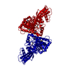
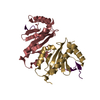
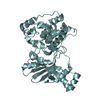
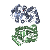
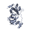
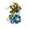
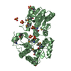
| Deposited unit | 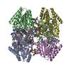
| ||||||||
|---|---|---|---|---|---|---|---|---|---|
| 1 | 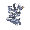
| ||||||||
| 2 | 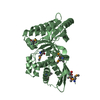
| ||||||||
| 3 | 
| ||||||||
| 4 | 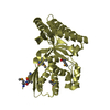
| ||||||||
| 5 | 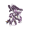
| ||||||||
| 6 | 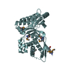
| ||||||||
| Unit cell |
| ||||||||
| Details | The protein is a monomer in solution and crystallizes as a hexameric asymetric unit of 3/222 noncrystallographic symmetry |
-Components
| #1: Protein | Mass: 37706.867 Da / Num. of mol.: 6 Source method: isolated from a genetically manipulated source Source: (gene. exp.) Streptococcus pneumoniae R6 (bacteria) / Species: Streptococcus pneumoniae / Strain: ATCC BAA-255 / R6 / Gene: mvaK2 / Plasmid: pGEX6P-1 / Species (production host): Escherichia coli / Production host: #2: Water | ChemComp-HOH / |  Mass: 18.015 Da / Num. of mol.: 779 / Source method: isolated from a natural source / Formula: H2O Mass: 18.015 Da / Num. of mol.: 779 / Source method: isolated from a natural source / Formula: H2OHas protein modification | Y | |
|---|
-Experimental details
-Experiment
| Experiment | Method: X-RAY DIFFRACTION / Number of used crystals: 1 |
|---|
- Sample preparation
| Crystal | Density Matthews: 3.14 Å3/Da / Density % sol: 60.5 % | |||||||||||||||||||||||||||||||||||||||||||||||||||||||||||||||
|---|---|---|---|---|---|---|---|---|---|---|---|---|---|---|---|---|---|---|---|---|---|---|---|---|---|---|---|---|---|---|---|---|---|---|---|---|---|---|---|---|---|---|---|---|---|---|---|---|---|---|---|---|---|---|---|---|---|---|---|---|---|---|---|---|
| Crystal grow | Temperature: 298 K / Method: vapor diffusion, hanging drop / pH: 7.5 Details: sodium dihydrogen phosphate, potassium dihydrogen phosphate, HEPES, ATP, pH 7.5, VAPOR DIFFUSION, HANGING DROP, temperature 298K | |||||||||||||||||||||||||||||||||||||||||||||||||||||||||||||||
| Crystal grow | *PLUS Temperature: 20 ℃ / pH: 7 | |||||||||||||||||||||||||||||||||||||||||||||||||||||||||||||||
| Components of the solutions | *PLUS
|
-Data collection
| Diffraction | Mean temperature: 100 K |
|---|---|
| Diffraction source | Source: SYNCHROTRON / Site: NSLS  / Beamline: X25 / Wavelength: 0.9792 Å / Beamline: X25 / Wavelength: 0.9792 Å |
| Detector | Type: BRANDEIS - B4 / Detector: CCD / Date: Feb 28, 2001 Details: 27-pole hybrid wiggler and vertical focusing mirror |
| Radiation | Monochromator: double Si crystal / Protocol: SINGLE WAVELENGTH / Monochromatic (M) / Laue (L): M / Scattering type: x-ray |
| Radiation wavelength | Wavelength: 0.9792 Å / Relative weight: 1 |
| Reflection | Resolution: 2.4→20 Å / Num. all: 202265 / Num. obs: 198856 / % possible obs: 98.3 % / Observed criterion σ(F): 1 / Observed criterion σ(I): 1 / Redundancy: 6.8 % / Biso Wilson estimate: 46 Å2 / Rmerge(I) obs: 0.082 / Rsym value: 0.082 / Net I/σ(I): 24.1 |
| Reflection shell | Resolution: 2.4→2.49 Å / Rmerge(I) obs: 0.409 / Mean I/σ(I) obs: 2.5 / Num. unique all: 18706 / Rsym value: 0.409 / % possible all: 92.3 |
| Reflection | *PLUS Highest resolution: 2.4 Å / Lowest resolution: 20 Å / Num. obs: 103387 / Num. measured all: 3616027 / Rmerge(I) obs: 0.082 |
| Reflection shell | *PLUS % possible obs: 92.4 % / Rmerge(I) obs: 0.409 |
- Processing
| Software |
| |||||||||||||||||||||||||
|---|---|---|---|---|---|---|---|---|---|---|---|---|---|---|---|---|---|---|---|---|---|---|---|---|---|---|
| Refinement | Method to determine structure: Single anomalous scattering / Resolution: 2.42→20 Å / Isotropic thermal model: anisotropic / Cross valid method: THROUGHOUT / σ(F): 0 / σ(I): 0 / Stereochemistry target values: Engh & Huber Details: anomalous scattering correction applied for 48 Se sites
| |||||||||||||||||||||||||
| Displacement parameters |
| |||||||||||||||||||||||||
| Refine analyze |
| |||||||||||||||||||||||||
| Refinement step | Cycle: LAST / Resolution: 2.42→20 Å
| |||||||||||||||||||||||||
| Refine LS restraints |
| |||||||||||||||||||||||||
| LS refinement shell |
| |||||||||||||||||||||||||
| Refinement | *PLUS Lowest resolution: 20 Å / % reflection Rfree: 9.2 % / Rfactor all: 0.217 / Rfactor obs: 0.215 / Rfactor Rfree: 0.257 / Rfactor Rwork: 0.215 | |||||||||||||||||||||||||
| Solvent computation | *PLUS | |||||||||||||||||||||||||
| Displacement parameters | *PLUS | |||||||||||||||||||||||||
| Refine LS restraints | *PLUS
| |||||||||||||||||||||||||
| LS refinement shell | *PLUS Rfactor Rfree: 0.312 / Rfactor Rwork: 0.305 / Rfactor obs: 0.305 |
