 Movie
Movie Controller

[English] 日本語
 Yorodumi
- PDB-2ib5: Structural characterization of a blue chromoprotein and its yello... -
+ Open data

- Basic information
| Entry | Database: PDB / ID: 2ib5 | ||||||||||||
|---|---|---|---|---|---|---|---|---|---|---|---|---|---|
| Title | Structural characterization of a blue chromoprotein and its yellow mutant from the sea anemone cnidopus japonicus | ||||||||||||
 Components Components | Chromo protein | ||||||||||||
 Keywords Keywords | LUMINESCENT PROTEIN / beta barrel / alpha helix / chromoprotein / chromophore / blue / GFP-like protein | ||||||||||||
| Function / homology |  Function and homology information Function and homology information | ||||||||||||
| Biological species |  Cnidopus japonicus (sea anemone) Cnidopus japonicus (sea anemone) | ||||||||||||
| Method |  X-RAY DIFFRACTION / SYNCHROTRON / SAD / Resolution: 1.8 Å X-RAY DIFFRACTION / SYNCHROTRON / SAD / Resolution: 1.8 Å | ||||||||||||
 Authors Authors | Chan, M.C.Y. / Bosanac, I. / Ikura, M. | ||||||||||||
 Citation Citation | Journal: J.Biol.Chem. / Year: 2006 Title: Structural Characterization of a Blue Chromoprotein and Its Yellow Mutant from the Sea Anemone Cnidopus Japonicus Authors: Chan, M.C.Y. / Karasawa, S. / Mizuno, H. / Bosanac, I. / Ho, D. / Prive, G.G. / Miyawaki, A. / Ikura, M. | ||||||||||||
| History |
|
- Structure visualization
| Structure viewer | Molecule: MolmilJmol/JSmol |
|---|
- Downloads & links
-Download
| PDBx/mmCIF format | 2ib5.cif.gz | 394.6 KB | Display | PDBx/mmCIF format |
|---|---|---|---|---|
| PDB format | pdb2ib5.ent.gz | 322.8 KB | Display | PDB format |
| PDBx/mmJSON format | 2ib5.json.gz | Tree view | PDBx/mmJSON format | |
| Others |  Other downloads Other downloads |
-Validation report
| Arichive directory | https://data.pdbj.org/pub/pdb/validation_reports/ib/2ib5ftp://data.pdbj.org/pub/pdb/validation_reports/ib/2ib5 | HTTPS FTP |
|---|
-Related structure data


-Links
 PDBj
PDBj

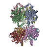
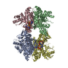
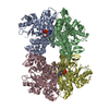
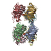
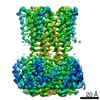
- Assembly
| Deposited unit | 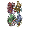
| ||||||||
|---|---|---|---|---|---|---|---|---|---|
| 1 |
| ||||||||
| 2 | 
| ||||||||
| 3 | 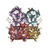
| ||||||||
| Unit cell |
| ||||||||
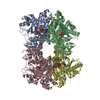
| Details | The biological assembly is a octomer generated from the tetramer in the asymmetric unit. |
-Components
| #1: Protein | Mass: 26842.227 Da / Num. of mol.: 8 Source method: isolated from a genetically manipulated source Source: (gene. exp.) Cnidopus japonicus (sea anemone) / Plasmid: pET28a / Production host:  #2: Chemical | ChemComp-PO4 /   Mass: 94.971 Da / Num. of mol.: 16 / Source method: obtained synthetically / Formula: PO4 Mass: 94.971 Da / Num. of mol.: 16 / Source method: obtained synthetically / Formula: PO4#3: Water | ChemComp-HOH / |  Mass: 18.015 Da / Num. of mol.: 1328 / Source method: isolated from a natural source / Formula: H2O Mass: 18.015 Da / Num. of mol.: 1328 / Source method: isolated from a natural source / Formula: H2OHas protein modification | Y | Sequence details | RESIDUES GLN 63, TYR 64 AND GLY 65 ARE MODIFIED TO FORM A CHROMOPHORE (CRQ 65) IN ALL CHAINS. ...RESIDUES GLN 63, TYR 64 AND GLY 65 ARE MODIFIED TO FORM A CHROMOPHOR | |
|---|
-Experimental details
-Experiment
| Experiment | Method: X-RAY DIFFRACTION / Number of used crystals: 1 |
|---|
- Sample preparation
| Crystal | Density Matthews: 2.14 Å3/Da / Density % sol: 42.6 % Description: THE STRUCTURE FACTOR FILE CONTAINS FRIEDEL PAIRS. |
|---|---|
| Crystal grow | Temperature: 295 K / Method: vapor diffusion, hanging drop / pH: 7.5 Details: 0.2M NaH2PO4, 20mM Tris-HCl, 150mM NaCl, 20% PEG3350, 20% glycerol, 2mM TCEP, pH 7.5, VAPOR DIFFUSION, HANGING DROP, temperature 295K |
-Data collection
| Diffraction | Mean temperature: 100 K |
|---|---|
| Diffraction source | Source: SYNCHROTRON / Site: APS  / Beamline: 19-ID / Wavelength: 0.9793 Å / Beamline: 19-ID / Wavelength: 0.9793 Å |
| Detector | Type: SBC-2 / Detector: CCD / Date: Dec 9, 2004 Details: Rosenbaum-Rock monochromator #1 high-resolution double-crystal sagittal focusing, Rosenbaum-Rock monochromator #2 double crystal, Rosenbaum-Rock vertical focusing mirror |
| Radiation | Monochromator: Rosenbaum-Rock double-crystal monochromator / Protocol: SINGLE WAVELENGTH / Monochromatic (M) / Laue (L): M / Scattering type: x-ray |
| Radiation wavelength | Wavelength: 0.9793 Å / Relative weight: 1 |
| Reflection | Resolution: 1.8→50 Å / Num. all: 335346 / Num. obs: 335346 / % possible obs: 99.3 % / Observed criterion σ(F): 0 / Observed criterion σ(I): 0 / Redundancy: 52.7 % / Biso Wilson estimate: 19.5 Å2 / Rmerge(I) obs: 0.077 / Net I/σ(I): 12 |
| Reflection shell | Resolution: 1.8→1.86 Å / Mean I/σ(I) obs: 5.1 / Num. unique all: 16336 / % possible all: 92 |
- Processing
| Software |
| |||||||||||||||||||||||||||
|---|---|---|---|---|---|---|---|---|---|---|---|---|---|---|---|---|---|---|---|---|---|---|---|---|---|---|---|---|
| Refinement | Method to determine structure: SAD / Resolution: 1.8→50 Å / Isotropic thermal model: anisotropic / Cross valid method: THROUGHOUT / σ(F): 0 / σ(I): 0 / Stereochemistry target values: Engh & Huber / Details: THE STRUCTURE FACTOR FILE CONTAINS FRIEDEL PAIRS.
| |||||||||||||||||||||||||||
| Displacement parameters | Biso mean: 19.5 Å2 | |||||||||||||||||||||||||||
| Refine analyze |
| |||||||||||||||||||||||||||
| Refinement step | Cycle: LAST / Resolution: 1.8→50 Å
| |||||||||||||||||||||||||||
| Refine LS restraints |
| |||||||||||||||||||||||||||
| LS refinement shell | Resolution: 1.8→1.86 Å / Rfactor Rfree error: 0.05
|
