 Movie
Movie Controller

[English] 日本語
 Yorodumi
- PDB-1xmz: Crystal structure of the dark state of kindling fluorescent prote... -
+ Open data

- Basic information
| Entry | Database: PDB / ID: 1xmz | |||||||||||||||
|---|---|---|---|---|---|---|---|---|---|---|---|---|---|---|---|---|
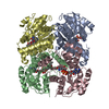
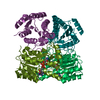
| Title | Crystal structure of the dark state of kindling fluorescent protein kfp from anemonia sulcata | |||||||||||||||
 Components Components |
| |||||||||||||||
 Keywords Keywords | LUMINESCENT PROTEIN / FLUORESCENT PROTEIN / CHROMOPHORE STRUCTURE / ASCP595 | |||||||||||||||
| Function / homology | Green fluorescent protein-related / Green fluorescent protein / Green fluorescent protein / bioluminescence / BETA-MERCAPTOETHANOL / GFP-like non-fluorescent chromoprotein FP595 Function and homology information Function and homology information | |||||||||||||||
| Biological species |  Anemonia sulcata (snake-locks sea anemone) Anemonia sulcata (snake-locks sea anemone) | |||||||||||||||
| Method |  X-RAY DIFFRACTION / SYNCHROTRON / MOLECULAR REPLACEMENT / Resolution: 1.38 Å X-RAY DIFFRACTION / SYNCHROTRON / MOLECULAR REPLACEMENT / Resolution: 1.38 Å | |||||||||||||||
 Authors Authors | Quillin, M.L. / Anstrom, D.M. / Shu, X. / O'Leary, S. / Kallio, K. / Chudakov, D.M. / Remington, S.J. | |||||||||||||||
 Citation Citation | Journal: Biochemistry / Year: 2005 Title: Kindling Fluorescent Protein from Anemonia sulcata: Dark-State Structure at 1.38 Resolution Authors: Quillin, M.L. / Anstrom, D.M. / Shu, X. / O'Leary, S. / Kallio, K. / Chudakov, D.M. / Remington, S.J. #1: Journal: J.Biol.Chem. / Year: 2000Title: Natural Animal Coloration Can be Determined by a Nonfluorescent Green Fluorescent Protein Homolog Authors: Lukyanov, K.A. / Fradkov, A.F. / Gurskaya, N.G. / Matz, M.V. / Labas, Y.A. / Savitsky, A.P. / Markelov, M.L. / Zaraisky, A.G. / Zhao, X. / Fang, Y. / Tan, W. / Lukyanov, S.A. #2: Journal: J.Biol.Chem. / Year: 2001Title: Alternative Cyclization in Gfp-Like Proteins Family. The Formation and Structure of the Chromophore of a Purple Chromoprotein from Anemonia Sulcata Authors: Martynov, V.I. / Savitsky, A.P. / Martynova, N.Y. / Savitsky, P.A. / Lukyanov, K.A. / Lukyanov, S.A. #3: Journal: BMC BIOCHEM. / Year: 2002Title: Interconversion of Anthozoa Gfp-Like Fluorescent and Nonfluorescent Proteins by Mutagenesis Authors: Bulina, M.E. / Chudakov, D.M. / Mudrik, N.N. / Lukyanov, K.A. #4: Journal: J.Biol.Chem. / Year: 2002Title: Chromophore Environment Provides Clue to "Kindling Fluorescent Protein" Riddle Authors: Chudakov, D.M. / Foefanov, A.V. / Mudrik, N.N. / Lukyanov, S. / Lukyanov, K.A. #5: Journal: NAT.BIOTECHNOL. / Year: 2003Title: Kindling Fluorescent Proteins for Precise in Vivo Photolabeling Authors: Chudakov, D.M. / Belousov, V.V. / Zaraisky, A.G. / Novoselov, V.V. / Staroverov, D.B. / Zorov, D.B. / Lukyanov, S. / Lukyanov, K.A. #6: Journal: To be PublishedTitle: Synthesis and Properties of the Chromophore of Asulcp Chromoprotein from Anemonia Sulcata Authors: Yampolsky, I.V. / Remington, S.J. / Potapov, V.K. / Lukyanov, K.A. | |||||||||||||||
| History |
| |||||||||||||||
| Remark 999 | SEQUENCE RESIDUE MET 63, TYR 64 AND GLY 65 ARE MODIFIED TO FORM A CHROMOPHORE (CRK 65) IN BOTH CHAINS | |||||||||||||||
| Remark 400 | COMPOUND THE AMINO GROUP (NH2 240) FOLLOWING CYS 62 IS DERIVED FROM THE PEPTIDE NITROGEN OF MET 63 |
- Structure visualization
| Structure viewer | Molecule: MolmilJmol/JSmol |
|---|
- Downloads & links
-Download
| PDBx/mmCIF format | 1xmz.cif.gz | 224 KB | Display | PDBx/mmCIF format |
|---|---|---|---|---|
| PDB format | pdb1xmz.ent.gz | 180.9 KB | Display | PDB format |
| PDBx/mmJSON format | 1xmz.json.gz | Tree view | PDBx/mmJSON format | |
| Others |  Other downloads Other downloads |
-Validation report
| Arichive directory | https://data.pdbj.org/pub/pdb/validation_reports/xm/1xmzftp://data.pdbj.org/pub/pdb/validation_reports/xm/1xmz | HTTPS FTP |
|---|
-Related structure data
| Related structure data |  1g7kS S: Starting model for refinement |
|---|---|
| Similar structure data |


-Links
 PDBj
PDBj


- Assembly
| Deposited unit | 
| ||||||||
|---|---|---|---|---|---|---|---|---|---|
| 1 | 
| ||||||||
| Unit cell |
| ||||||||
| Noncrystallographic symmetry (NCS) | NCS oper: (Code: given Matrix: (0.188266, 0.002404, -0.982115), Vector: |
-Components
| #1: Protein | Mass: 8065.189 Da / Num. of mol.: 2 Source method: isolated from a genetically manipulated source Source: (gene. exp.) Anemonia sulcata (snake-locks sea anemone)Plasmid: PQE-30 / Production host:  #2: Protein | Mass: 19143.678 Da / Num. of mol.: 2 / Mutation: A143G Source method: isolated from a genetically manipulated source Source: (gene. exp.) Anemonia sulcata (snake-locks sea anemone)Plasmid: PQE-30 / Production host: #3: Chemical | ChemComp-BME /   Mass: 78.133 Da / Num. of mol.: 7 / Source method: obtained synthetically / Formula: C2H6OS Mass: 78.133 Da / Num. of mol.: 7 / Source method: obtained synthetically / Formula: C2H6OS#4: Water | ChemComp-HOH / |  Mass: 18.015 Da / Num. of mol.: 463 / Source method: isolated from a natural source / Formula: H2O Mass: 18.015 Da / Num. of mol.: 463 / Source method: isolated from a natural source / Formula: H2OHas protein modification | Y | |
|---|
-Experimental details
-Experiment
| Experiment | Method: X-RAY DIFFRACTION / Number of used crystals: 1 |
|---|
- Sample preparation
| Crystal | Density Matthews: 2.05 Å3/Da / Density % sol: 39.9 % |
|---|---|
| Crystal grow | Temperature: 293 K / Method: vapor diffusion, hanging drop / pH: 9.5 Details: 26% PEG 1550, 0.14 M SODIUM CITRATE, 0.1 M TRIS, pH 9.5, VAPOR DIFFUSION, HANGING DROP, temperature 293K |
-Data collection
| Diffraction | Mean temperature: 100 K |
|---|---|
| Diffraction source | Source: SYNCHROTRON / Site: APS  / Beamline: 14-BM-C / Wavelength: 0.9 / Wavelength: 0.9 Å / Beamline: 14-BM-C / Wavelength: 0.9 / Wavelength: 0.9 Å |
| Detector | Type: ADSC QUANTUM 4 / Detector: CCD / Date: Nov 12, 2003 |
| Radiation | Protocol: SINGLE WAVELENGTH / Monochromatic (M) / Laue (L): M / Scattering type: x-ray |
| Radiation wavelength | Wavelength: 0.9 Å / Relative weight: 1 |
| Reflection | Resolution: 1.38→50 Å / Num. all: 89428 / Num. obs: 89428 / % possible obs: 97.5 % / Observed criterion σ(F): 0 / Observed criterion σ(I): 0 / Redundancy: 7.3 % / Biso Wilson estimate: 14.9 Å2 / Rmerge(I) obs: 0.044 / Net I/σ(I): 49.1 |
| Reflection shell | Resolution: 1.38→1.43 Å / Rmerge(I) obs: 0.166 / Mean I/σ(I) obs: 8.4 / Num. unique all: 8895 / % possible all: 97.9 |
- Processing
| Software |
| |||||||||||||||||||||||||||||||||
|---|---|---|---|---|---|---|---|---|---|---|---|---|---|---|---|---|---|---|---|---|---|---|---|---|---|---|---|---|---|---|---|---|---|---|
| Refinement | Method to determine structure: MOLECULAR REPLACEMENT Starting model: PDB ENTRY 1G7K Resolution: 1.38→50 Å / Num. parameters: 38699 / Num. restraintsaints: 48588 / Isotropic thermal model: INDIVIDUAL ANISOTROPIC B-FACTORS / Cross valid method: FREE R / σ(F): 0 / Stereochemistry target values: Engh & Huber
| |||||||||||||||||||||||||||||||||
| Solvent computation | Solvent model: A solvent model based on Moews & Kretsinger, 1975 was applied during refinement. | |||||||||||||||||||||||||||||||||
| Refine analyze | Num. disordered residues: 46 / Occupancy sum hydrogen: 3322 / Occupancy sum non hydrogen: 4108 | |||||||||||||||||||||||||||||||||
| Refinement step | Cycle: LAST / Resolution: 1.38→50 Å
| |||||||||||||||||||||||||||||||||
| Refine LS restraints |
|
