 Movie
Movie Controller


+ Open data

- Basic information
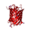
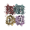
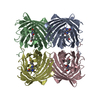
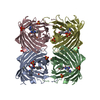
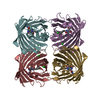
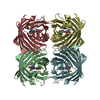
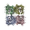
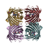
| Entry | Database: PDB / ID: 1mou | ||||||||||||
|---|---|---|---|---|---|---|---|---|---|---|---|---|---|
| Title | Crystal structure of Coral pigment | ||||||||||||
 Components Components | GFP-like non-fluorescent chromoprotein | ||||||||||||
 Keywords Keywords | LUMINESCENT PROTEIN / blue coral pigment / chromophore / beta can fold / similar to Green fluorescent protein and DsRed | ||||||||||||
| Function / homology |  Function and homology information Function and homology information | ||||||||||||
| Biological species |  Montipora efflorescens (invertebrata) Montipora efflorescens (invertebrata) | ||||||||||||
| Method |  X-RAY DIFFRACTION / MOLECULAR REPLACEMENT / Resolution: 2.2 Å X-RAY DIFFRACTION / MOLECULAR REPLACEMENT / Resolution: 2.2 Å | ||||||||||||
 Authors Authors | Prescott, M. / Ling, M. / Beddoe, T. / Oakley, A.J. / Dove, S. / Hoegh-Guldberg, O. / Devenish, R.J. / Rossjohn, J. | ||||||||||||
 Citation Citation | Journal: Structure / Year: 2003 Title: The 2.2 a crystal structure of a pocilloporin pigment reveals a nonplanar chromophore conformation. Authors: Prescott, M. / Ling, M. / Beddoe, T. / Oakley, A.J. / Dove, S. / Hoegh-Guldberg, O. / Devenish, R.J. / Rossjohn, J. | ||||||||||||
| History |
|
- Structure visualization
| Structure viewer | Molecule: MolmilJmol/JSmol |
|---|
- Downloads & links
-Download
| PDBx/mmCIF format | 1mou.cif.gz | 64.4 KB | Display | PDBx/mmCIF format |
|---|---|---|---|---|
| PDB format | pdb1mou.ent.gz | 45.3 KB | Display | PDB format |
| PDBx/mmJSON format | 1mou.json.gz | Tree view | PDBx/mmJSON format | |
| Others |  Other downloads Other downloads |
-Validation report
| Arichive directory | https://data.pdbj.org/pub/pdb/validation_reports/mo/1mouftp://data.pdbj.org/pub/pdb/validation_reports/mo/1mou | HTTPS FTP |
|---|
-Related structure data
| Related structure data |  1movC  1ggxS C: citing same article ( S: Starting model for refinement |
|---|---|
| Similar structure data |


-Links
 PDBj
PDBj

- Assembly
| Deposited unit | 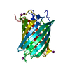
| ||||||||
|---|---|---|---|---|---|---|---|---|---|
| 1 | 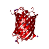
| ||||||||
| Unit cell |
| ||||||||
| Components on special symmetry positions |
|
-Components
| #1: Protein | Mass: 24815.123 Da / Num. of mol.: 1 / Fragment: RESIDUES 5-225 Source method: isolated from a genetically manipulated source Source: (gene. exp.) Montipora efflorescens (invertebrata) / Production host:  | ||||
|---|---|---|---|---|---|
| #2: Chemical | ChemComp-IOD /   Mass: 126.904 Da / Num. of mol.: 5 / Source method: obtained synthetically / Formula: I Mass: 126.904 Da / Num. of mol.: 5 / Source method: obtained synthetically / Formula: I#3: Water | ChemComp-HOH / |  Mass: 18.015 Da / Num. of mol.: 220 / Source method: isolated from a natural source / Formula: H2O Mass: 18.015 Da / Num. of mol.: 220 / Source method: isolated from a natural source / Formula: H2OHas protein modification | Y | |
-Experimental details
-Experiment
| Experiment | Method: X-RAY DIFFRACTION / Number of used crystals: 1 |
|---|
- Sample preparation
| Crystal grow | Temperature: 293 K / Method: vapor diffusion, hanging drop / pH: 8 Details: PEG, Tris buffer, pH 8, VAPOR DIFFUSION, HANGING DROP, temperature 293K | ||||||||||||||||||||||||||||||
|---|---|---|---|---|---|---|---|---|---|---|---|---|---|---|---|---|---|---|---|---|---|---|---|---|---|---|---|---|---|---|---|
| Crystal grow | *PLUS pH: 8.2 | ||||||||||||||||||||||||||||||
| Components of the solutions | *PLUS
|
-Data collection
| Diffraction | Mean temperature: 100 K |
|---|---|
| Diffraction source | Source: ROTATING ANODE / Type: RIGAKU RU300 / Wavelength: 1.54 Å |
| Detector | Type: RIGAKU RAXIS IV / Detector: IMAGE PLATE / Date: May 1, 2002 / Details: osmic mirrors |
| Radiation | Monochromator: none / Protocol: SINGLE WAVELENGTH / Monochromatic (M) / Laue (L): M / Scattering type: x-ray |
| Radiation wavelength | Wavelength: 1.54 Å / Relative weight: 1 |
| Reflection | Resolution: 2.2→100 Å / Num. all: 32494 / Num. obs: 32494 / % possible obs: 96.1 % / Observed criterion σ(F): -3 / Observed criterion σ(I): -3 / Redundancy: 3.4 % / Rmerge(I) obs: 0.079 / Rsym value: 0.079 / Net I/σ(I): 6.4 |
| Reflection shell | Resolution: 2.2→2.3 Å / Rmerge(I) obs: 0.247 / Mean I/σ(I) obs: 2.3 / Rsym value: 0.247 / % possible all: 98.5 |
| Reflection | *PLUS Lowest resolution: 100 Å / Num. measured all: 109869 |
| Reflection shell | *PLUS % possible obs: 98.5 % |
- Processing
| Software |
| |||||||||||||||||||||||||
|---|---|---|---|---|---|---|---|---|---|---|---|---|---|---|---|---|---|---|---|---|---|---|---|---|---|---|
| Refinement | Method to determine structure: MOLECULAR REPLACEMENT Starting model: 1ggx Resolution: 2.2→50 Å / Cross valid method: THROUGHOUT / σ(F): 0 / σ(I): 0
| |||||||||||||||||||||||||
| Refinement step | Cycle: LAST / Resolution: 2.2→50 Å
| |||||||||||||||||||||||||
| Refinement | *PLUS Lowest resolution: 50 Å / % reflection Rfree: 3 % | |||||||||||||||||||||||||
| Solvent computation | *PLUS | |||||||||||||||||||||||||
| Displacement parameters | *PLUS | |||||||||||||||||||||||||
| Refine LS restraints | *PLUS
|
