 Movie
Movie Controller


+ Open data

- Basic information
| Entry | Database: PDB / ID: 2h66 | ||||||
|---|---|---|---|---|---|---|---|
| Title | The Crystal Structure of Plasmodium Vivax 2-Cys peroxiredoxin | ||||||
 Components Components | PV-PF14_0368 | ||||||
 Keywords Keywords | STRUCTURAL GENOMICS/OXIDOREDUCTASE / plasmodium / vivax / peroxiredoxin / Structural Genomics / Structural Genomics Consortium / SGC / STRUCTURAL GENOMICS-OXIDOREDUCTASE COMPLEX | ||||||
| Function / homology |  Function and homology information Function and homology informationthioredoxin peroxidase activity / cellular response to stress / cell redox homeostasis / hydrogen peroxide catabolic process / response to oxidative stress / cytosol Similarity search - Function | ||||||
| Biological species |  | ||||||
| Method |  X-RAY DIFFRACTION / SYNCHROTRON / MOLECULAR REPLACEMENT / Resolution: 2.5 Å X-RAY DIFFRACTION / SYNCHROTRON / MOLECULAR REPLACEMENT / Resolution: 2.5 Å | ||||||
 Authors Authors | Wernimont, A.K. / Dong, A. / Zhao, Y. / Lew, J. / Melone, M. / Kozieradzki, I. / Weigelt, J. / Sundstrom, M. / Edwards, A.M. / Arrowsmith, C.H. ...Wernimont, A.K. / Dong, A. / Zhao, Y. / Lew, J. / Melone, M. / Kozieradzki, I. / Weigelt, J. / Sundstrom, M. / Edwards, A.M. / Arrowsmith, C.H. / Bochkarev, A. / Hui, R. / Artz, J.D. / Structural Genomics Consortium (SGC) | ||||||
 Citation Citation | Journal: Mol.Biochem.Parasitol. / Year: 2007 Title: Genome-scale protein expression and structural biology of Plasmodium falciparum and related Apicomplexan organisms. Authors: Vedadi, M. / Lew, J. / Artz, J. / Amani, M. / Zhao, Y. / Dong, A. / Wasney, G.A. / Gao, M. / Hills, T. / Brokx, S. / Qiu, W. / Sharma, S. / Diassiti, A. / Alam, Z. / Melone, M. / Mulichak, A. ...Authors: Vedadi, M. / Lew, J. / Artz, J. / Amani, M. / Zhao, Y. / Dong, A. / Wasney, G.A. / Gao, M. / Hills, T. / Brokx, S. / Qiu, W. / Sharma, S. / Diassiti, A. / Alam, Z. / Melone, M. / Mulichak, A. / Wernimont, A. / Bray, J. / Loppnau, P. / Plotnikova, O. / Newberry, K. / Sundararajan, E. / Houston, S. / Walker, J. / Tempel, W. / Bochkarev, A. / Kozieradzki, I. / Edwards, A. / Arrowsmith, C. / Roos, D. / Kain, K. / Hui, R. | ||||||
| History |
| ||||||
| Remark 999 | SEQUENCE Currently, there is no aminoacid sequence database reference available for the protein |
- Structure visualization
| Structure viewer | Molecule: MolmilJmol/JSmol |
|---|
- Downloads & links
-Download
| PDBx/mmCIF format | 2h66.cif.gz | 332.6 KB | Display | PDBx/mmCIF format |
|---|---|---|---|---|
| PDB format | pdb2h66.ent.gz | 271.3 KB | Display | PDB format |
| PDBx/mmJSON format | 2h66.json.gz | Tree view | PDBx/mmJSON format | |
| Others |  Other downloads Other downloads |
-Validation report
| Arichive directory | https://data.pdbj.org/pub/pdb/validation_reports/h6/2h66ftp://data.pdbj.org/pub/pdb/validation_reports/h6/2h66 | HTTPS FTP |
|---|
-Related structure data
| Related structure data | 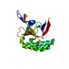 1txjC 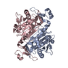 1xccC 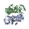 1y6zC 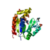 1z6gC 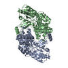 1z7dC 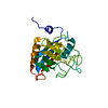 1z81C  1zo2C 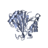 2a22C 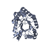 2a4aC 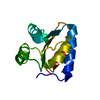 2aifC 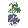 2amxC 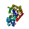 2aqwC 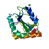 2av4C 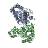 2awpC 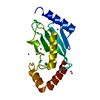 2ayvC 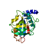 2b71C 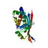 2bddC 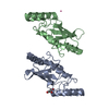 2f4zC 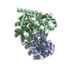 2fdsC 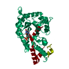 2ffcC 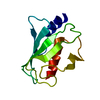 2fo3C 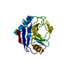 2fu0C 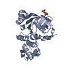 2ghiC 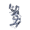 2h1rC 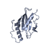 2h2yC 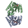 2hjrC 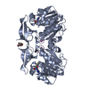 2hteC 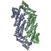 2hvgC  3pggC 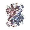 3tb2C  1qmvS S: Starting model for refinement C: citing same article ( |
|---|---|
| Similar structure data |


-Links
 PDBj
PDBj- Assembly
| Deposited unit | 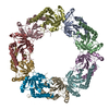
| ||||||||
|---|---|---|---|---|---|---|---|---|---|
| 1 |
| ||||||||
| Unit cell |
| ||||||||
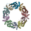
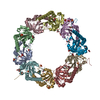
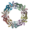
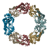
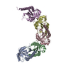
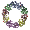
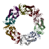
| Details | The biological assembly is a pentagonal ring of dimers, as seen in the asymmetric unit. |
-Components
| #1: Protein | Mass: 23732.133 Da / Num. of mol.: 10 Source method: isolated from a genetically manipulated source Source: (gene. exp.) Plasmid: pET28 LIC/TEV DER / Production host:  #2: Water | ChemComp-HOH / |  Mass: 18.015 Da / Num. of mol.: 154 / Source method: isolated from a natural source / Formula: H2O Mass: 18.015 Da / Num. of mol.: 154 / Source method: isolated from a natural source / Formula: H2OHas protein modification | Y | |
|---|
-Experimental details
-Experiment
| Experiment | Method: X-RAY DIFFRACTION / Number of used crystals: 2 |
|---|
- Sample preparation
| Crystal | Density Matthews: 2.83 Å3/Da / Density % sol: 56.59 % |
|---|---|
| Crystal grow | Temperature: 300 K / Method: vapor diffusion, hanging drop / pH: 4.6 Details: 5% PEG 4000, 50 mM NaAc, 100 mM NaAC, pH 4.6, VAPOR DIFFUSION, HANGING DROP, temperature 300K |
-Data collection
| Diffraction | Mean temperature: 100 K |
|---|---|
| Diffraction source | Source: SYNCHROTRON / Site: APS  / Beamline: 17-ID / Wavelength: 1 Å / Beamline: 17-ID / Wavelength: 1 Å |
| Detector | Type: ADSC QUANTUM 210 / Detector: CCD / Date: Mar 20, 2006 |
| Radiation | Protocol: SINGLE WAVELENGTH / Monochromatic (M) / Laue (L): M / Scattering type: x-ray |
| Radiation wavelength | Wavelength: 1 Å / Relative weight: 1 |
| Reflection | Resolution: 2.48→41.56 Å / Num. all: 91258 / Num. obs: 86441 / % possible obs: 99.2 % / Observed criterion σ(F): 0 / Observed criterion σ(I): 0 / Redundancy: 3.4 % / Biso Wilson estimate: 54.3 Å2 / Rmerge(I) obs: 0.115 / Rsym value: 0.098 / Net I/σ(I): 10.8 |
| Reflection shell | Resolution: 2.48→2.53 Å / Redundancy: 3.3 % / Rmerge(I) obs: 0.488 / Mean I/σ(I) obs: 1.3 / Num. unique all: 5688 / Rsym value: 0.498 / % possible all: 99.8 |
- Processing
| Software |
| |||||||||||||||||||||||||
|---|---|---|---|---|---|---|---|---|---|---|---|---|---|---|---|---|---|---|---|---|---|---|---|---|---|---|
| Refinement | Method to determine structure: MOLECULAR REPLACEMENT Starting model: PDB ENTRY 1QMV Resolution: 2.5→41.56 Å / Cross valid method: THROUGHOUT / σ(F): 0 / Stereochemistry target values: MAXIMUM LIKELIHOOD
| |||||||||||||||||||||||||
| Displacement parameters | Biso mean: 45.744 Å2
| |||||||||||||||||||||||||
| Refinement step | Cycle: LAST / Resolution: 2.5→41.56 Å
| |||||||||||||||||||||||||
| LS refinement shell | Resolution: 2.5→2.565 Å
|
