 Movie
Movie Controller

[English] 日本語
 Yorodumi
- PDB-1zo2: Structure of nuclear transport factor 2 (Ntf2) from Cryptosporidi... -
+ Open data

- Basic information
| Entry | Database: PDB / ID: 1zo2 | ||||||
|---|---|---|---|---|---|---|---|
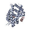
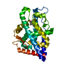
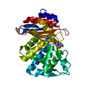
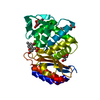
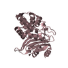
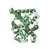
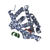
| Title | Structure of nuclear transport factor 2 (Ntf2) from Cryptosporidium parvum | ||||||
 Components Components | nuclear transport factor 2 | ||||||
 Keywords Keywords | TRANSPORT PROTEIN / NUCLEAR TRANSPORT / Structural Genomics / Structural Genomics Consortium / SGC | ||||||
| Function / homology |  Function and homology information Function and homology informationmRNA transport / protein import into nucleus / nuclear envelope / cytoplasm Similarity search - Function | ||||||
| Biological species |   Cryptosporidium parvum (eukaryote) Cryptosporidium parvum (eukaryote) | ||||||
| Method |  X-RAY DIFFRACTION / MOLECULAR REPLACEMENT / Resolution: 1.6 Å X-RAY DIFFRACTION / MOLECULAR REPLACEMENT / Resolution: 1.6 Å | ||||||
 Authors Authors | Choe, J. / Artz, J.D. / Gao, M. / Lew, J. / Zhao, Y. / Arrowsmith, C. / Edwards, A. / Sundstrom, M. / Bochkarev, A. / Hui, R. / Structural Genomics Consortium (SGC) | ||||||
 Citation Citation | Journal: Mol.Biochem.Parasitol. / Year: 2007 Title: Genome-scale protein expression and structural biology of Plasmodium falciparum and related Apicomplexan organisms. Authors: Vedadi, M. / Lew, J. / Artz, J. / Amani, M. / Zhao, Y. / Dong, A. / Wasney, G.A. / Gao, M. / Hills, T. / Brokx, S. / Qiu, W. / Sharma, S. / Diassiti, A. / Alam, Z. / Melone, M. / Mulichak, A. ...Authors: Vedadi, M. / Lew, J. / Artz, J. / Amani, M. / Zhao, Y. / Dong, A. / Wasney, G.A. / Gao, M. / Hills, T. / Brokx, S. / Qiu, W. / Sharma, S. / Diassiti, A. / Alam, Z. / Melone, M. / Mulichak, A. / Wernimont, A. / Bray, J. / Loppnau, P. / Plotnikova, O. / Newberry, K. / Sundararajan, E. / Houston, S. / Walker, J. / Tempel, W. / Bochkarev, A. / Kozieradzki, I. / Edwards, A. / Arrowsmith, C. / Roos, D. / Kain, K. / Hui, R. | ||||||
| History |
|
- Structure visualization
| Structure viewer | Molecule: MolmilJmol/JSmol |
|---|
- Downloads & links
-Download
| PDBx/mmCIF format | 1zo2.cif.gz | 64.7 KB | Display | PDBx/mmCIF format |
|---|---|---|---|---|
| PDB format | pdb1zo2.ent.gz | 47.2 KB | Display | PDB format |
| PDBx/mmJSON format | 1zo2.json.gz | Tree view | PDBx/mmJSON format | |
| Others |  Other downloads Other downloads |
-Validation report
| Arichive directory | https://data.pdbj.org/pub/pdb/validation_reports/zo/1zo2ftp://data.pdbj.org/pub/pdb/validation_reports/zo/1zo2 | HTTPS FTP |
|---|
-Related structure data
| Related structure data | 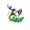 1txjC 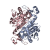 1xccC 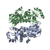 1y6zC 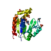 1z6gC  1z7dC  1z81C  2a22C  2a4aC 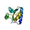 2aifC 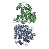 2amxC 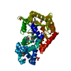 2aqwC 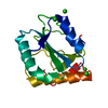 2av4C 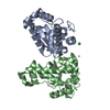 2awpC 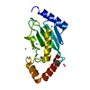 2ayvC 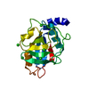 2b71C 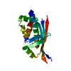 2bddC 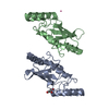 2f4zC 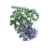 2fdsC 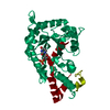 2ffcC 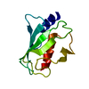 2fo3C 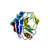 2fu0C 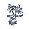 2ghiC 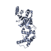 2h1rC 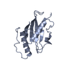 2h2yC  2h66C  2hjrC  2hteC  2hvgC  3pggC 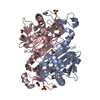 3tb2C  1gybS S: Starting model for refinement C: citing same article ( |
|---|---|
| Similar structure data |


-Links
 PDBj
PDBj
- Assembly
| Deposited unit | 
| ||||||||
|---|---|---|---|---|---|---|---|---|---|
| 1 |
| ||||||||
| Unit cell |
|
-Components
| #1: Protein | Mass: 14698.395 Da / Num. of mol.: 2 Source method: isolated from a genetically manipulated source Source: (gene. exp.) Cryptosporidium parvum (eukaryote) / Plasmid: pET28 / Species (production host): Escherichia coli / Production host:  #2: Water | ChemComp-HOH / |  Mass: 18.015 Da / Num. of mol.: 178 / Source method: isolated from a natural source / Formula: H2O Mass: 18.015 Da / Num. of mol.: 178 / Source method: isolated from a natural source / Formula: H2O |
|---|
-Experimental details
-Experiment
| Experiment | Method: X-RAY DIFFRACTION / Number of used crystals: 1 |
|---|
- Sample preparation
| Crystal | Density Matthews: 2.1 Å3/Da / Density % sol: 41.7 % |
|---|---|
| Crystal grow | Temperature: 291 K / Method: vapor diffusion, sitting drop / pH: 7.5 Details: PEG 3350, Ammonium phosphate, pH 7.5, VAPOR DIFFUSION, SITTING DROP, temperature 291K |
-Data collection
| Diffraction | Mean temperature: 100 K |
|---|---|
| Diffraction source | Source: ROTATING ANODE / Type: RIGAKU FR-E / Wavelength: 1.5418 Å |
| Detector | Type: RIGAKU RAXIS IV / Detector: IMAGE PLATE / Date: May 11, 2005 |
| Radiation | Monochromator: CONFOCAL MAXFLUX OPTICS / Protocol: SINGLE WAVELENGTH / Monochromatic (M) / Laue (L): M / Scattering type: x-ray |
| Radiation wavelength | Wavelength: 1.5418 Å / Relative weight: 1 |
| Reflection | Resolution: 1.6→35.3 Å / Num. all: 28649 / Num. obs: 28649 / % possible obs: 91.8 % / Observed criterion σ(F): 0 / Observed criterion σ(I): 0 / Redundancy: 6.7 % / Biso Wilson estimate: 22.5 Å2 / Rmerge(I) obs: 0.046 / Net I/σ(I): 35.7 |
| Reflection shell | Resolution: 1.6→1.66 Å / Redundancy: 4.2 % / Rmerge(I) obs: 0.269 / Mean I/σ(I) obs: 5.2 / % possible all: 56.2 |
- Processing
| Software |
| ||||||||||||||||||||||||||||||||||||||||||||||||||||||||||||
|---|---|---|---|---|---|---|---|---|---|---|---|---|---|---|---|---|---|---|---|---|---|---|---|---|---|---|---|---|---|---|---|---|---|---|---|---|---|---|---|---|---|---|---|---|---|---|---|---|---|---|---|---|---|---|---|---|---|---|---|---|---|
| Refinement | Method to determine structure: MOLECULAR REPLACEMENT Starting model: PDb entry 1GYB Resolution: 1.6→35.16 Å / Rfactor Rfree error: 0.007 / Data cutoff high absF: 823352.82 / Data cutoff low absF: 0 / Isotropic thermal model: RESTRAINED / Cross valid method: THROUGHOUT / σ(F): 0 / Stereochemistry target values: Engh & Huber
| ||||||||||||||||||||||||||||||||||||||||||||||||||||||||||||
| Solvent computation | Solvent model: FLAT MODEL / Bsol: 51.8368 Å2 / ksol: 0.404979 e/Å3 | ||||||||||||||||||||||||||||||||||||||||||||||||||||||||||||
| Displacement parameters | Biso mean: 19.3 Å2
| ||||||||||||||||||||||||||||||||||||||||||||||||||||||||||||
| Refine analyze |
| ||||||||||||||||||||||||||||||||||||||||||||||||||||||||||||
| Refinement step | Cycle: LAST / Resolution: 1.6→35.16 Å
| ||||||||||||||||||||||||||||||||||||||||||||||||||||||||||||
| Refine LS restraints |
| ||||||||||||||||||||||||||||||||||||||||||||||||||||||||||||
| LS refinement shell | Resolution: 1.6→1.7 Å / Rfactor Rfree error: 0.028 / Total num. of bins used: 6
| ||||||||||||||||||||||||||||||||||||||||||||||||||||||||||||
| Xplor file |
|
