 Movie
Movie Controller


+ Open data

- Basic information
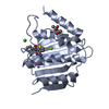
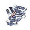
| Entry | Database: PDB / ID: 1z6g | ||||||
|---|---|---|---|---|---|---|---|
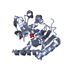
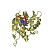
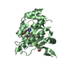
| Title | Crystal structure of guanylate kinase from Plasmodium falciparum | ||||||
 Components Components | guanylate kinase | ||||||
 Keywords Keywords | TRANSFERASE / guanylate kinase / Structural Genomics / SGC / Structural Genomics Consortium | ||||||
| Function / homology |  Function and homology information Function and homology informationAzathioprine ADME / Interconversion of nucleotide di- and triphosphates / guanylate kinase / GMP kinase activity / ATP binding / cytosol Similarity search - Function | ||||||
| Biological species |  | ||||||
| Method |  X-RAY DIFFRACTION / SYNCHROTRON / MOLECULAR REPLACEMENT / Resolution: 2.18 Å X-RAY DIFFRACTION / SYNCHROTRON / MOLECULAR REPLACEMENT / Resolution: 2.18 Å | ||||||
 Authors Authors | Mulichak, A.M. / Lew, J. / Artz, J. / Choe, J. / Walker, J.R. / Zhao, Y. / Sundstrom, M. / Arrowsmith, C. / Edwards, A. / Bochkarev, A. ...Mulichak, A.M. / Lew, J. / Artz, J. / Choe, J. / Walker, J.R. / Zhao, Y. / Sundstrom, M. / Arrowsmith, C. / Edwards, A. / Bochkarev, A. / Hui, R. / Gao, M. / Structural Genomics Consortium (SGC) | ||||||
 Citation Citation | Journal: Mol.Biochem.Parasitol. / Year: 2007 Title: Genome-scale protein expression and structural biology of Plasmodium falciparum and related Apicomplexan organisms. Authors: Vedadi, M. / Lew, J. / Artz, J. / Amani, M. / Zhao, Y. / Dong, A. / Wasney, G.A. / Gao, M. / Hills, T. / Brokx, S. / Qiu, W. / Sharma, S. / Diassiti, A. / Alam, Z. / Melone, M. / Mulichak, A. ...Authors: Vedadi, M. / Lew, J. / Artz, J. / Amani, M. / Zhao, Y. / Dong, A. / Wasney, G.A. / Gao, M. / Hills, T. / Brokx, S. / Qiu, W. / Sharma, S. / Diassiti, A. / Alam, Z. / Melone, M. / Mulichak, A. / Wernimont, A. / Bray, J. / Loppnau, P. / Plotnikova, O. / Newberry, K. / Sundararajan, E. / Houston, S. / Walker, J. / Tempel, W. / Bochkarev, A. / Kozieradzki, I. / Edwards, A. / Arrowsmith, C. / Roos, D. / Kain, K. / Hui, R. | ||||||
| History |
|
- Structure visualization
| Structure viewer | Molecule: MolmilJmol/JSmol |
|---|
- Downloads & links
-Download
| PDBx/mmCIF format | 1z6g.cif.gz | 57.2 KB | Display | PDBx/mmCIF format |
|---|---|---|---|---|
| PDB format | pdb1z6g.ent.gz | 39.9 KB | Display | PDB format |
| PDBx/mmJSON format | 1z6g.json.gz | Tree view | PDBx/mmJSON format | |
| Others |  Other downloads Other downloads |
-Validation report
| Arichive directory | https://data.pdbj.org/pub/pdb/validation_reports/z6/1z6gftp://data.pdbj.org/pub/pdb/validation_reports/z6/1z6g | HTTPS FTP |
|---|
-Related structure data
| Related structure data |  1txjC  1xccC  1y6zC  1z7dC 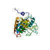 1z81C  1zo2C  2a22C  2a4aC 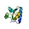 2aifC 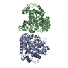 2amxC 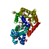 2aqwC 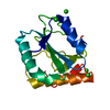 2av4C 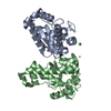 2awpC 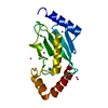 2ayvC 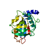 2b71C 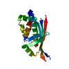 2bddC 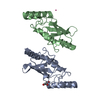 2f4zC  2fdsC 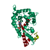 2ffcC 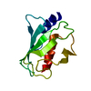 2fo3C 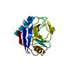 2fu0C 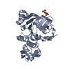 2ghiC 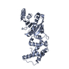 2h1rC 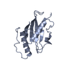 2h2yC 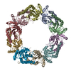 2h66C 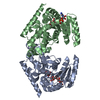 2hjrC 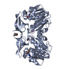 2hteC 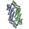 2hvgC  3pggC 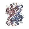 3tb2C  1ex7S 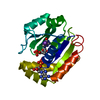 1lvgS S: Starting model for refinement C: citing same article ( |
|---|---|
| Similar structure data |


-Links
 PDBj
PDBj- Assembly
| Deposited unit | 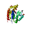
| ||||||||
|---|---|---|---|---|---|---|---|---|---|
| 1 |
| ||||||||
| Unit cell |
|
-Components
| #1: Protein | Mass: 25579.088 Da / Num. of mol.: 1 Source method: isolated from a genetically manipulated source Source: (gene. exp.) Strain: 3D7 / Plasmid: custom pET-28a / Production host:  | ||||
|---|---|---|---|---|---|
| #2: Chemical | ChemComp-SO4 /   Mass: 96.063 Da / Num. of mol.: 5 / Source method: obtained synthetically / Formula: SO4 Mass: 96.063 Da / Num. of mol.: 5 / Source method: obtained synthetically / Formula: SO4#3: Chemical | ChemComp-EPE / |   Mass: 238.305 Da / Num. of mol.: 1 / Source method: obtained synthetically / Formula: C8H18N2O4S / Comment: pH buffer*YM Mass: 238.305 Da / Num. of mol.: 1 / Source method: obtained synthetically / Formula: C8H18N2O4S / Comment: pH buffer*YM#4: Water | ChemComp-HOH / |  Mass: 18.015 Da / Num. of mol.: 131 / Source method: isolated from a natural source / Formula: H2O Mass: 18.015 Da / Num. of mol.: 131 / Source method: isolated from a natural source / Formula: H2O |
-Experimental details
-Experiment
| Experiment | Method: X-RAY DIFFRACTION / Number of used crystals: 1 |
|---|
- Sample preparation
| Crystal | Density Matthews: 2.46 Å3/Da / Density % sol: 49.53 % |
|---|---|
| Crystal grow | Temperature: 298 K / Method: vapor diffusion / pH: 7.4 Details: ammonium sulfate, hepes buffer, pH 7.4, VAPOR DIFFUSION, temperature 298K |
-Data collection
| Diffraction | Mean temperature: 100 K |
|---|---|
| Diffraction source | Source: SYNCHROTRON / Site: APS  / Beamline: 17-ID / Wavelength: 1 Å / Beamline: 17-ID / Wavelength: 1 Å |
| Detector | Type: ADSC QUANTUM 210 / Detector: CCD / Date: Feb 6, 2005 |
| Radiation | Monochromator: Si 111 / Protocol: SINGLE WAVELENGTH / Monochromatic (M) / Laue (L): M / Scattering type: x-ray |
| Radiation wavelength | Wavelength: 1 Å / Relative weight: 1 |
| Reflection | Resolution: 2.1→50 Å / Num. all: 14255 / Num. obs: 13877 / % possible obs: 99.8 % / Observed criterion σ(F): 0 / Observed criterion σ(I): 0 / Redundancy: 5 % / Rsym value: 0.061 |
| Reflection shell | Resolution: 2.1→2.18 Å / Redundancy: 5 % / Num. unique all: 1401 / Rsym value: 0.211 / % possible all: 100 |
- Processing
| Software |
| ||||||||||||||||||||
|---|---|---|---|---|---|---|---|---|---|---|---|---|---|---|---|---|---|---|---|---|---|
| Refinement | Method to determine structure: MOLECULAR REPLACEMENT Starting model: PDB entries 1EX7 and 1LVG Resolution: 2.18→30 Å / Cross valid method: THROUGHOUT / σ(F): 0 / Stereochemistry target values: Engh & Huber
| ||||||||||||||||||||
| Refinement step | Cycle: LAST / Resolution: 2.18→30 Å
|
