 Movie
Movie Controller


+ Open data

- Basic information
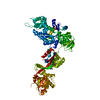
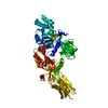
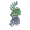
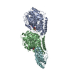
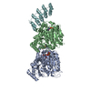
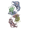
| Entry | Database: PDB / ID: 1n0v | ||||||
|---|---|---|---|---|---|---|---|
| Title | Crystal structure of elongation factor 2 | ||||||
 Components Components | Elongation factor 2 | ||||||
 Keywords Keywords | TRANSLATION / G-protein cis-proline | ||||||
| Function / homology |  Function and homology information Function and homology informationPeptide chain elongation / Synthesis of diphthamide-EEF2 / positive regulation of translational elongation / Protein methylation / translational elongation / translation elongation factor activity / Neutrophil degranulation / maintenance of translational fidelity / protein-folding chaperone binding / ribosome binding ...Peptide chain elongation / Synthesis of diphthamide-EEF2 / positive regulation of translational elongation / Protein methylation / translational elongation / translation elongation factor activity / Neutrophil degranulation / maintenance of translational fidelity / protein-folding chaperone binding / ribosome binding / Hydrolases; Acting on acid anhydrides; Acting on GTP to facilitate cellular and subcellular movement / rRNA binding / ribonucleoprotein complex / GTPase activity / GTP binding / identical protein binding / cytosol Similarity search - Function | ||||||
| Biological species |  | ||||||
| Method |  X-RAY DIFFRACTION / SYNCHROTRON / MOLECULAR REPLACEMENT / Resolution: 2.85 Å X-RAY DIFFRACTION / SYNCHROTRON / MOLECULAR REPLACEMENT / Resolution: 2.85 Å | ||||||
 Authors Authors | Joergensen, R. / Ortiz, P.A. / Carr-Schmid, A. / Nissen, P. / Kinzy, T.G. / Andersen, G.R. | ||||||
 Citation Citation | Journal: Nat.Struct.Biol. / Year: 2003 Title: Two crystal structures demonstrate large conformational changes in the eukaryotic ribosomal translocase. Authors: Joergensen, R. / Ortiz, P.A. / Carr-Schmid, A. / Nissen, P. / Kinzy, T.G. / Andersen, G.R. #1: Journal: Acta Crystallogr.,Sect.D / Year: 2002Title: Purification and Crystallization of the yeast elongation factor eEF2 Authors: Joergensen, R. / Carr-Schmid, A. / Ortiz, P.A. / Kinzy, T.G. / Andersen, G.R. | ||||||
| History |
|
- Structure visualization
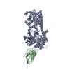
| Structure viewer | Molecule: MolmilJmol/JSmol |
|---|
- Downloads & links
-Download
| PDBx/mmCIF format | 1n0v.cif.gz | 321.2 KB | Display | PDBx/mmCIF format |
|---|---|---|---|---|
| PDB format | pdb1n0v.ent.gz | 260.8 KB | Display | PDB format |
| PDBx/mmJSON format | 1n0v.json.gz | Tree view | PDBx/mmJSON format | |
| Others |  Other downloads Other downloads |
-Validation report
| Arichive directory | https://data.pdbj.org/pub/pdb/validation_reports/n0/1n0vftp://data.pdbj.org/pub/pdb/validation_reports/n0/1n0v | HTTPS FTP |
|---|
-Related structure data


-Links
 PDBj
PDBj


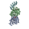
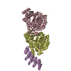
- Assembly
| Deposited unit | 
| ||||||||
|---|---|---|---|---|---|---|---|---|---|
| 1 | 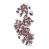
| ||||||||
| 2 | 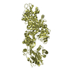
| ||||||||
| Unit cell |
| ||||||||
| Details | The molecule functions as a monomer |
-Components
| #1: Protein | Mass: 93407.125 Da / Num. of mol.: 2 / Source method: isolated from a natural source / Source: (natural) |
|---|
-Experimental details
-Experiment
| Experiment | Method: X-RAY DIFFRACTION / Number of used crystals: 1 |
|---|
- Sample preparation
| Crystal | Density Matthews: 2.69 Å3/Da / Density % sol: 54.21 % | |||||||||||||||||||||||||||||||||||||||||||||||||||||||||||||||||||||||||||||||||||||||||||
|---|---|---|---|---|---|---|---|---|---|---|---|---|---|---|---|---|---|---|---|---|---|---|---|---|---|---|---|---|---|---|---|---|---|---|---|---|---|---|---|---|---|---|---|---|---|---|---|---|---|---|---|---|---|---|---|---|---|---|---|---|---|---|---|---|---|---|---|---|---|---|---|---|---|---|---|---|---|---|---|---|---|---|---|---|---|---|---|---|---|---|---|---|
| Crystal grow | Method: vapor diffusion / pH: 7.2 Details: PEG8K, Hepes, ethylene glycol; magnesium chloride, GDP, EDTA, DTT, pH 7.2, VAPOR DIFFUSION, temperature 100K | |||||||||||||||||||||||||||||||||||||||||||||||||||||||||||||||||||||||||||||||||||||||||||
| Crystal grow | *PLUS Temperature: 277 K / Method: vapor diffusion, sitting dropDetails: Joergensen, R., (2002) Acta Crystallogr., Sect.D, 58, 712. | |||||||||||||||||||||||||||||||||||||||||||||||||||||||||||||||||||||||||||||||||||||||||||
| Components of the solutions | *PLUS
|
-Data collection
| Diffraction | Mean temperature: 100 K |
|---|---|
| Diffraction source | Source: SYNCHROTRON / Site: MAX II  / Beamline: I711 / Wavelength: 1.038 Å / Beamline: I711 / Wavelength: 1.038 Å |
| Detector | Type: MARRESEARCH / Detector: CCD / Date: Nov 8, 2001 |
| Radiation | Protocol: SINGLE WAVELENGTH / Monochromatic (M) / Laue (L): M / Scattering type: x-ray |
| Radiation wavelength | Wavelength: 1.038 Å / Relative weight: 1 |
| Reflection | Resolution: 2.85→30 Å / Num. all: 47695 / Num. obs: 45917 / % possible obs: 96.3 % / Observed criterion σ(F): 0 / Observed criterion σ(I): 0 / Redundancy: 5.3 % / Rmerge(I) obs: 0.054 / Net I/σ(I): 39.1 |
| Reflection shell | Resolution: 2.85→2.95 Å / Rmerge(I) obs: 0.316 / % possible all: 97 |
| Reflection | *PLUS Lowest resolution: 30 Å |
- Processing
| Software |
| ||||||||||||||||||||
|---|---|---|---|---|---|---|---|---|---|---|---|---|---|---|---|---|---|---|---|---|---|
| Refinement | Method to determine structure: MOLECULAR REPLACEMENT Starting model: Structure of eEF2-sordarin complex Resolution: 2.85→30 Å / Isotropic thermal model: isotropic / Cross valid method: THROUGHOUT / σ(F): 0 / Stereochemistry target values: Engh & Huber
| ||||||||||||||||||||
| Refinement step | Cycle: LAST / Resolution: 2.85→30 Å
| ||||||||||||||||||||
| Refine LS restraints |
| ||||||||||||||||||||
| Refinement | *PLUS Lowest resolution: 30 Å | ||||||||||||||||||||
| Solvent computation | *PLUS | ||||||||||||||||||||
| Displacement parameters | *PLUS |
