 Movie
Movie Controller


+ Open data

- Basic information
| Entry | Database: PDB / ID: 1fnm | ||||||
|---|---|---|---|---|---|---|---|
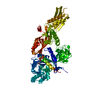
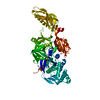
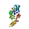
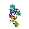
| Title | STRUCTURE OF THERMUS THERMOPHILUS EF-G H573A | ||||||
 Components Components | ELONGATION FACTOR G | ||||||
 Keywords Keywords | TRANSLATION / Bent conformation / Visible domain III / Mutation His573Ala | ||||||
| Function / homology |  Function and homology information Function and homology informationribosome disassembly / translational elongation / translation elongation factor activity / GDP binding / ribosome binding / GTPase activity / GTP binding / magnesium ion binding / cytoplasm Similarity search - Function | ||||||
| Biological species |   Thermus thermophilus (bacteria) Thermus thermophilus (bacteria) | ||||||
| Method |  X-RAY DIFFRACTION / SYNCHROTRON / Resolution: 2.8 Å X-RAY DIFFRACTION / SYNCHROTRON / Resolution: 2.8 Å | ||||||
 Authors Authors | Laurberg, M. / Kristensen, O. / Martemyanov, K. / Gudkov, A.T. / Nagaev, I. / Hughes, D. / Liljas, A. | ||||||
 Citation Citation | Journal: J.Mol.Biol. / Year: 2000 Title: Structure of a mutant EF-G reveals domain III and possibly the fusidic acid binding site. Authors: Laurberg, M. / Kristensen, O. / Martemyanov, K. / Gudkov, A.T. / Nagaev, I. / Hughes, D. / Liljas, A. #1: Journal: Embo J. / Year: 1994Title: The Crystal Structure of Elongation Factor G complexed with GDP, at 2.7 A Resolution Authors: Czworkowski, J. / Wang, J. / Steitz, T.A. / Moore, P.B. #2: Journal: Embo J. / Year: 1994Title: Three-dimensional Structure of the Ribosomal Translocase: Elongation Factor G from Thermus thermophilus Authors: Aevarsson, A. / Brazhnikov, E. / Garber, M. / Zheltonosova, J. / Chirgadze, Y. / al-Karadaghi, S. / Svensson, L.A. / Liljas, A. #3: Journal: Structure / Year: 1996Title: The Structure of Elongation Factor G in Complex with GDP: Conformational Flexibility and Nucleotide Exchange Authors: al-Karadaghi, S. / Aevarsson, A. / Garber, M. / Zheltonosova, J. / Liljas, A. #4: Journal: FEBS Lett. / Year: 1998Title: An Intact Conformation at the Tip of Elongation Factor G Domain IV is Functionally Important Authors: Martemyanov, K.A. / Yarunin, A.S. / Liljas, A. / Gudkov, A.T. | ||||||
| History |
|
- Structure visualization
| Structure viewer | Molecule: MolmilJmol/JSmol |
|---|
- Downloads & links
-Download
| PDBx/mmCIF format | 1fnm.cif.gz | 129.8 KB | Display | PDBx/mmCIF format |
|---|---|---|---|---|
| PDB format | pdb1fnm.ent.gz | 101.5 KB | Display | PDB format |
| PDBx/mmJSON format | 1fnm.json.gz | Tree view | PDBx/mmJSON format | |
| Others |  Other downloads Other downloads |
-Validation report
| Arichive directory | https://data.pdbj.org/pub/pdb/validation_reports/fn/1fnmftp://data.pdbj.org/pub/pdb/validation_reports/fn/1fnm | HTTPS FTP |
|---|
-Related structure data
| Similar structure data |
|---|


-Links
 PDBj
PDBj


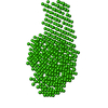
- Assembly
| Deposited unit | 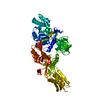
| ||||||||
|---|---|---|---|---|---|---|---|---|---|
| 1 |
| ||||||||
| Unit cell |
|
-Components
| #1: Protein | Mass: 76910.031 Da / Num. of mol.: 1 / Mutation: H573A Source method: isolated from a genetically manipulated source Source: (gene. exp.) Thermus thermophilus (bacteria) / Plasmid: PET11C (NOVAGEN) / Production host: |
|---|---|
| #2: Chemical | ChemComp-MG /   Mass: 24.305 Da / Num. of mol.: 1 / Source method: obtained synthetically / Formula: Mg Mass: 24.305 Da / Num. of mol.: 1 / Source method: obtained synthetically / Formula: Mg |
| #3: Chemical | ChemComp-GDP /   Type: RNA linking / Mass: 443.201 Da / Num. of mol.: 1 / Source method: obtained synthetically / Formula: C10H15N5O11P2 / Comment: GDP, energy-carrying molecule*YM Type: RNA linking / Mass: 443.201 Da / Num. of mol.: 1 / Source method: obtained synthetically / Formula: C10H15N5O11P2 / Comment: GDP, energy-carrying molecule*YM |
| #4: Water | ChemComp-HOH /  Mass: 18.015 Da / Num. of mol.: 18 / Source method: isolated from a natural source / Formula: H2O Mass: 18.015 Da / Num. of mol.: 18 / Source method: isolated from a natural source / Formula: H2O |
-Experimental details
-Experiment
| Experiment | Method: X-RAY DIFFRACTION / Number of used crystals: 1 |
|---|
- Sample preparation
| Crystal | Density Matthews: 2.43 Å3/Da / Density % sol: 49.35 % | ||||||||||||||||||||||||||||||||||||||||||||||||
|---|---|---|---|---|---|---|---|---|---|---|---|---|---|---|---|---|---|---|---|---|---|---|---|---|---|---|---|---|---|---|---|---|---|---|---|---|---|---|---|---|---|---|---|---|---|---|---|---|---|
| Crystal grow | Temperature: 293 K / Method: vapor diffusion, hanging drop / pH: 7.6 Details: PEG 8000 (Fluka), guanosine 5'-diphophate, magnesium chloride , pH 7.6, VAPOR DIFFUSION, HANGING DROP, temperature 293.0K | ||||||||||||||||||||||||||||||||||||||||||||||||
| Crystal grow | *PLUS | ||||||||||||||||||||||||||||||||||||||||||||||||
| Components of the solutions | *PLUS
|
-Data collection
| Diffraction | Mean temperature: 100 K |
|---|---|
| Diffraction source | Source: SYNCHROTRON / Site: MAX II  / Beamline: I711 / Wavelength: 0.935 / Beamline: I711 / Wavelength: 0.935 |
| Detector | Type: MARRESEARCH / Detector: IMAGE PLATE / Date: May 12, 1998 |
| Radiation | Protocol: SINGLE WAVELENGTH / Monochromatic (M) / Laue (L): M / Scattering type: x-ray |
| Radiation wavelength | Wavelength: 0.935 Å / Relative weight: 1 |
| Reflection | Resolution: 2.8→25 Å / Num. all: 18060 / Num. obs: 18060 / % possible obs: 94.8 % / Observed criterion σ(F): -4 / Observed criterion σ(I): -3 / Redundancy: 4.2 % / Biso Wilson estimate: 44.8 Å2 / Rmerge(I) obs: 0.1 / Net I/σ(I): 12.5 |
| Reflection shell | Resolution: 2.8→2.9 Å / Redundancy: 4.2 % / Rmerge(I) obs: 0.41 / Num. unique all: 2011 / % possible all: 96.8 |
| Reflection | *PLUS Rmerge(I) obs: 0.104 |
| Reflection shell | *PLUS % possible obs: 96.8 % |
- Processing
| Software |
| |||||||||||||||||||||||||
|---|---|---|---|---|---|---|---|---|---|---|---|---|---|---|---|---|---|---|---|---|---|---|---|---|---|---|
| Refinement | Resolution: 2.8→25 Å / σ(F): 0 / σ(I): 0 / Stereochemistry target values: Engh & Huber
| |||||||||||||||||||||||||
| Refinement step | Cycle: LAST / Resolution: 2.8→25 Å
| |||||||||||||||||||||||||
| Refine LS restraints |
| |||||||||||||||||||||||||
| Software | *PLUS Name: CNS / Classification: refinement | |||||||||||||||||||||||||
| Refinement | *PLUS Highest resolution: 2.8 Å / Lowest resolution: 25 Å / σ(F): 0 / % reflection Rfree: 3.6 % / Rfactor obs: 0.212 | |||||||||||||||||||||||||
| Solvent computation | *PLUS | |||||||||||||||||||||||||
| Displacement parameters | *PLUS | |||||||||||||||||||||||||
| Refine LS restraints | *PLUS
|
