 Movie
Movie Controller


+ Open data

- Basic information
| Entry | Database: PDB / ID: 2j7k | ||||||
|---|---|---|---|---|---|---|---|
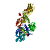
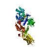
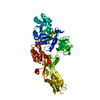
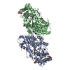
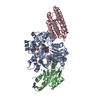
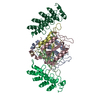
| Title | Crystal structure of the T84A mutant EF-G:GDPCP complex | ||||||
 Components Components | ELONGATION FACTOR G | ||||||
 Keywords Keywords | TRANSLATION / ELONGATION FACTOR / NUCLEOTIDE-BINDING / P-LOOP / THR84ALA / MUTATION / GTP-BINDING / NUCLEOTIDE- BINDING / PROTEIN BIOSYNTHESIS | ||||||
| Function / homology |  Function and homology information Function and homology informationribosome disassembly / translational elongation / translation elongation factor activity / GDP binding / ribosome binding / GTPase activity / GTP binding / magnesium ion binding / cytoplasm Similarity search - Function | ||||||
| Biological species |   THERMUS THERMOPHILUS (bacteria) THERMUS THERMOPHILUS (bacteria) | ||||||
| Method |  X-RAY DIFFRACTION / SYNCHROTRON / MOLECULAR REPLACEMENT / Resolution: 2.9 Å X-RAY DIFFRACTION / SYNCHROTRON / MOLECULAR REPLACEMENT / Resolution: 2.9 Å | ||||||
 Authors Authors | Hansson, S. / Logan, D.T. | ||||||
 Citation Citation | Journal: To be Published Title: New Insights Into the Role of the P-Loop Lysine: Implications from the Crystal Structure of a Mutant EF-G:Gdpcp Complex Authors: Hansson, S. / Logan, D.T. #1: Journal: FEBS Lett. / Year: 2005Title: Crystal Structure of a Mutant Elongation Factor G Trapped with a GTP Analogue Authors: Hansson, S. / Singh, R. / Gudkov, A.T. / Liljas, A. / Logan, D.T. | ||||||
| History |
| ||||||
| Remark 700 | SHEET DETERMINATION METHOD: DSSP THE SHEETS PRESENTED AS "AD" IN EACH CHAIN ON SHEET RECORDS BELOW ... SHEET DETERMINATION METHOD: DSSP THE SHEETS PRESENTED AS "AD" IN EACH CHAIN ON SHEET RECORDS BELOW IS ACTUALLY AN 7-STRANDED BARREL THIS IS REPRESENTED BY A 8-STRANDED SHEET IN WHICH THE FIRST AND LAST STRANDS ARE IDENTICAL. THE SHEET STRUCTURE OF THIS MOLECULE IS BIFURCATED. IN ORDER TO REPRESENT THIS FEATURE IN THE SHEET RECORDS BELOW, TWO SHEETS ARE DEFINED. |
- Structure visualization
| Structure viewer | Molecule: MolmilJmol/JSmol |
|---|
- Downloads & links
-Download
| PDBx/mmCIF format | 2j7k.cif.gz | 141.8 KB | Display | PDBx/mmCIF format |
|---|---|---|---|---|
| PDB format | pdb2j7k.ent.gz | 109.4 KB | Display | PDB format |
| PDBx/mmJSON format | 2j7k.json.gz | Tree view | PDBx/mmJSON format | |
| Others |  Other downloads Other downloads |
-Validation report
| Arichive directory | https://data.pdbj.org/pub/pdb/validation_reports/j7/2j7kftp://data.pdbj.org/pub/pdb/validation_reports/j7/2j7k | HTTPS FTP |
|---|
-Related structure data
| Related structure data | 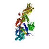 2bm0S S: Starting model for refinement |
|---|---|
| Similar structure data |


-Links
 PDBj
PDBj


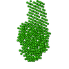
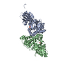
- Assembly
| Deposited unit | 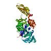
| ||||||||
|---|---|---|---|---|---|---|---|---|---|
| 1 |
| ||||||||
| Unit cell |
|
-Components
| #1: Protein | Mass: 76947.078 Da / Num. of mol.: 1 / Mutation: YES Source method: isolated from a genetically manipulated source Source: (gene. exp.) THERMUS THERMOPHILUS (bacteria) / Strain: HB8 / Plasmid: PET28 / Production host: |
|---|---|
| #2: Chemical | ChemComp-GCP /   Mass: 521.208 Da / Num. of mol.: 1 / Source method: obtained synthetically / Formula: C11H18N5O13P3 / Comment: GMP-PCP, energy-carrying molecule analogue*YM Mass: 521.208 Da / Num. of mol.: 1 / Source method: obtained synthetically / Formula: C11H18N5O13P3 / Comment: GMP-PCP, energy-carrying molecule analogue*YM |
| #3: Chemical | ChemComp-MG /   Mass: 24.305 Da / Num. of mol.: 1 / Source method: obtained synthetically / Formula: Mg Mass: 24.305 Da / Num. of mol.: 1 / Source method: obtained synthetically / Formula: Mg |
| #4: Water | ChemComp-HOH /  Mass: 18.015 Da / Num. of mol.: 3 / Source method: isolated from a natural source / Formula: H2O Mass: 18.015 Da / Num. of mol.: 3 / Source method: isolated from a natural source / Formula: H2O |
| Compound details | ENGINEERED |
-Experimental details
-Experiment
| Experiment | Method: X-RAY DIFFRACTION |
|---|
- Sample preparation
| Crystal | Density Matthews: 2.7 Å3/Da / Density % sol: 54.11 % / Description: NONE |
|---|---|
| Crystal grow | pH: 7.3 Details: 17 % PEG8000 100 MM HEPES 46 MM TRIS 10 MM MGCL2 10 MM GDPCP, pH 7.3 |
-Data collection
| Diffraction | Mean temperature: 100 K |
|---|---|
| Diffraction source | Source: SYNCHROTRON / Site: ESRF  / Beamline: ID29 / Wavelength: 1.065 / Beamline: ID29 / Wavelength: 1.065 |
| Detector | Type: ADSC CCD / Detector: CCD / Date: Feb 21, 2005 / Details: MIRRORS |
| Radiation | Protocol: SINGLE WAVELENGTH / Monochromatic (M) / Laue (L): M / Scattering type: x-ray |
| Radiation wavelength | Wavelength: 1.065 Å / Relative weight: 1 |
| Reflection | Resolution: 2.9→26 Å / Num. obs: 17014 / % possible obs: 95.4 % / Observed criterion σ(I): 2.5 / Redundancy: 7.4 % / Rmerge(I) obs: 0.07 / Net I/σ(I): 18.6 |
| Reflection shell | Resolution: 2.9→3 Å / Rmerge(I) obs: 0.47 / Mean I/σ(I) obs: 3.2 / % possible all: 98.7 |
- Processing
| Software |
| ||||||||||||||||||||
|---|---|---|---|---|---|---|---|---|---|---|---|---|---|---|---|---|---|---|---|---|---|
| Refinement | Method to determine structure: MOLECULAR REPLACEMENT Starting model: PDB ENTRY 2BM0 Resolution: 2.9→26 Å / Cross valid method: THROUGHOUT / Stereochemistry target values: MAXIMUM LIKELIHOOD Details: HYDROGENS HAVE BEEN ADDED IN THE RIDING POSITIONS. PEPTIDE BOND BETWEEN RESIDUES 85 AND 86 IS A CIS-PEPTIDE.
| ||||||||||||||||||||
| Displacement parameters | Biso mean: 67.74 Å2 | ||||||||||||||||||||
| Refinement step | Cycle: LAST / Resolution: 2.9→26 Å
| ||||||||||||||||||||
| LS refinement shell | Resolution: 2.9→2.97 Å / Num. reflection Rwork: 1679 |
