[English] 日本語
Yorodumi
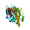
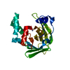
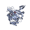
- PDB-5ttz: Crystal structure of Grp94 bound to isoform-selective inhibitor m... -

+
Open data


ID or keywords:

Loading...

-
Basic information

Entry
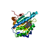
Database: PDB / ID: 5ttz
Title
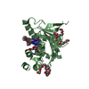
Crystal structure of Grp94 bound to isoform-selective inhibitor methyl 2-(2-(1-(4-bromobenzyl)-1H-imidazol-2-yl)ethyl)-3-chloro-4,6-dihydroxybenzoate
ComponentsEndoplasmin
KeywordsCHAPERONE / INHIBITOR / BnIm Scaffold / ATP Binding Site / CHAPERONE - INHIBITOR complex
Function / homology
Function and homology information


Trafficking and processing of endosomal TLR / Scavenging by Class A Receptors / Regulation of Insulin-like Growth Factor (IGF) transport and uptake by Insulin-like Growth Factor Binding Proteins (IGFBPs) / Interleukin-4 and Interleukin-13 signaling / Post-translational protein phosphorylation / sarcoplasmic reticulum lumen / ERAD pathway / ATP-dependent protein folding chaperone / unfolded protein binding / melanosome ...Trafficking and processing of endosomal TLR / Scavenging by Class A Receptors / Regulation of Insulin-like Growth Factor (IGF) transport and uptake by Insulin-like Growth Factor Binding Proteins (IGFBPs) / Interleukin-4 and Interleukin-13 signaling / Post-translational protein phosphorylation / sarcoplasmic reticulum lumen / ERAD pathway / ATP-dependent protein folding chaperone / unfolded protein binding / melanosome / protein folding / response to heat / DNA damage response / perinuclear region of cytoplasm / endoplasmic reticulum / ATP hydrolysis activity / ATP binding / cytosol
Similarity search - Function
Endoplasmic reticulum targeting sequence. / Heat shock protein Hsp90, conserved site / Heat shock hsp90 proteins family signature. / HSP90, C-terminal domain / Heat shock protein Hsp90, N-terminal / Heat shock protein Hsp90 family / Hsp90 protein / Histidine kinase-, DNA gyrase B-, and HSP90-like ATPase / Histidine kinase-like ATPase, C-terminal domain / Heat Shock Protein 90 ...Endoplasmic reticulum targeting sequence. / Heat shock protein Hsp90, conserved site / Heat shock hsp90 proteins family signature. / HSP90, C-terminal domain / Heat shock protein Hsp90, N-terminal / Heat shock protein Hsp90 family / Hsp90 protein / Histidine kinase-, DNA gyrase B-, and HSP90-like ATPase / Histidine kinase-like ATPase, C-terminal domain / Heat Shock Protein 90 / Histidine kinase-, DNA gyrase B-, and HSP90-like ATPase / Histidine kinase-like ATPases / Histidine kinase/HSP90-like ATPase / Histidine kinase/HSP90-like ATPase superfamily / Ribosomal protein S5 domain 2-type fold / 2-Layer Sandwich / Alpha Beta
Similarity search - Domain/homology
Chem-7KY / DI(HYDROXYETHYL)ETHER / TRIETHYLENE GLYCOL / Endoplasmin
Similarity search - Component
Biological speciesCanis lupus familiaris (dog)
MethodX-RAY DIFFRACTION / SYNCHROTRON / MOLECULAR REPLACEMENT / Resolution: 2.707 Å
AuthorsLieberman, R.L. / Huard, D.J.E.
Funding support United States, 1items
OrganizationGrant numberCountry
National Institutes of Health/National Eye Institute (NIH/NEI)R01EY021205 United States
CitationJournal: Sci Rep / Year: 2017
Title: Isoform-selective Hsp90 inhibition rescues model of hereditary open-angle glaucoma.
Authors: Stothert, A.R. / Suntharalingam, A. / Tang, X. / Crowley, V.M. / Mishra, S.J. / Webster, J.M. / Nordhues, B.A. / Huard, D.J.E. / Passaglia, C.L. / Lieberman, R.L. / Blagg, B.S.J. / Blair, L. ...Authors: Stothert, A.R. / Suntharalingam, A. / Tang, X. / Crowley, V.M. / Mishra, S.J. / Webster, J.M. / Nordhues, B.A. / Huard, D.J.E. / Passaglia, C.L. / Lieberman, R.L. / Blagg, B.S.J. / Blair, L.J. / Koren, J. / Dickey, C.A.
History
DepositionNov 4, 2016Deposition site: RCSB / Processing site: RCSB
Revision 1.0Jan 10, 2018Provider: repository / Type: Initial release
Revision 1.1Dec 11, 2019Group: Author supporting evidence / Category: pdbx_audit_support / Item: _pdbx_audit_support.funding_organization
Revision 1.2Oct 4, 2023Group: Advisory / Data collection ...Advisory / Data collection / Database references / Refinement description
Category: chem_comp_atom / chem_comp_bond ...chem_comp_atom / chem_comp_bond / database_2 / pdbx_initial_refinement_model / pdbx_unobs_or_zero_occ_atoms
Item: _database_2.pdbx_DOI / _database_2.pdbx_database_accession

-
Structure visualization

Structure viewerMolecule:
MolmilJmol/JSmol

Downloads & links

-
Assembly

Deposited unit
A: Endoplasmin
B: Endoplasmin
hetero molecules


Theoretical massNumber of molelcules
Total (without water)54,1128
Polymers52,3752
Non-polymers1,7366
Water66737
1
A: Endoplasmin
hetero molecules


Theoretical massNumber of molelcules
Total (without water)26,7593
Polymers26,1881
Non-polymers5722
Water181
TypeNameSymmetry operationNumber
identity operation1_555x,y,z1
MethodPISA
2
B: Endoplasmin
hetero molecules


Theoretical massNumber of molelcules
Total (without water)27,3525
Polymers26,1881
Non-polymers1,1654
Water181
TypeNameSymmetry operationNumber
identity operation1_555x,y,z1
MethodPISA
Unit cell
Length a, b, c (Å)65.237, 84.420, 95.473
Angle α, β, γ (deg.)90.00, 90.00, 90.00
Int Tables number19
Space group name H-MP212121

-
Components

-
Protein , 1 types, 2 molecules AB

#1: Protein Endoplasmin / 94 kDa glucose-regulated protein / GRP-94 / Heat shock protein 90 kDa beta member 1


Mass: 26187.615 Da / Num. of mol.: 2 / Fragment: residues 69-337
Source method: isolated from a genetically manipulated source
Source: (gene. exp.) Canis lupus familiaris (dog) / Gene: HSP90B1, GRP94, TRA1 / Production host: Escherichia coli (E. coli) / Strain (production host): BL21(DE3) / References: UniProt: P41148

-
Non-polymers , 6 types, 43 molecules

#2: Chemical ChemComp-7KY / methyl 2-(2-{1-[(4-bromophenyl)methyl]-1H-imidazol-2-yl}ethyl)-3-chloro-4,6-dihydroxybenzoate


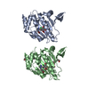
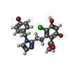
Mass: 465.725 Da / Num. of mol.: 2 / Source method: obtained synthetically / Formula: C20H18BrClN2O4
#3: Chemical ChemComp-PEG / DI(HYDROXYETHYL)ETHER


Mass: 106.120 Da / Num. of mol.: 1 / Source method: obtained synthetically / Formula: C4H10O3
#4: Chemical ChemComp-PG4 / TETRAETHYLENE GLYCOL


Mass: 194.226 Da / Num. of mol.: 1 / Source method: obtained synthetically / Formula: C8H18O5 / Comment: precipitant*YM
#5: Chemical ChemComp-PGE / TRIETHYLENE GLYCOL


Mass: 150.173 Da / Num. of mol.: 1 / Source method: obtained synthetically / Formula: C6H14O4
#6: Chemical ChemComp-PE4 / 2-{2-[2-(2-{2-[2-(2-ETHOXY-ETHOXY)-ETHOXY]-ETHOXY}-ETHOXY)-ETHOXY]-ETHOXY}-ETHANOL / POLYETHYLENE GLYCOL PEG4000


Mass: 354.436 Da / Num. of mol.: 1 / Source method: obtained synthetically / Formula: C16H34O8 / Comment: precipitant*YM
#7: Water ChemComp-HOH / water


Mass: 18.015 Da / Num. of mol.: 37 / Source method: isolated from a natural source / Formula: H2O

-
Experimental details

-
Experiment

ExperimentMethod: X-RAY DIFFRACTION / Number of used crystals: 1

-
Sample preparation

CrystalDensity Matthews: 2.51 Å3/Da / Density % sol: 50.99 %
Crystal growTemperature: 289.15 K / Method: vapor diffusion, hanging drop / pH: 7.5
Details: Protein at 30 mg/mL in 0.1 M Tris pH 7.5 was mixed 1:1 with mother liquor comprised of 35% PEG400, 0.1 M Tris pH 7.5, and 80 mM MgCl2. Crystals were harvested and soaked in mother liquor ...Details: Protein at 30 mg/mL in 0.1 M Tris pH 7.5 was mixed 1:1 with mother liquor comprised of 35% PEG400, 0.1 M Tris pH 7.5, and 80 mM MgCl2. Crystals were harvested and soaked in mother liquor containing 20 mM inhibitor. A layer of glycerol was then added through which crystals were harvested and cryo-cooled in liquid nitrogen.

-
Data collection

DiffractionMean temperature: 80 K
Diffraction sourceSource: SYNCHROTRON / Site: APS / Beamline: 22-BM / Wavelength: 1 Å
DetectorType: MARMOSAIC 300 mm CCD / Detector: CCD / Date: Jun 25, 2014
RadiationProtocol: SINGLE WAVELENGTH / Monochromatic (M) / Laue (L): M / Scattering type: x-ray
Radiation wavelengthWavelength: 1 Å / Relative weight: 1
ReflectionResolution: 2.707→47.74 Å / Num. obs: 217928 / % possible obs: 97.71 % / Redundancy: 14.6 % / Rmerge(I) obs: 0.121 / Net I/σ(I): 22.11
Reflection shellRmerge(I) obs: 0.663

-
Processing

Software
NameVersionClassification
PHENIX1.9_1692refinement
XDSdata reduction
XSCALEdata scaling
PHASERphasing
RefinementMethod to determine structure: MOLECULAR REPLACEMENT
Starting model: 2GFD
Resolution: 2.707→47.736 Å / SU ML: 0.42 / Cross valid method: FREE R-VALUE / σ(F): 1.35 / Phase error: 31.51
RfactorNum. reflection% reflection
Rfree0.2855 1453 10 %
Rwork0.1992 --
obs0.2076 14528 97.4 %
Solvent computationShrinkage radii: 0.9 Å / VDW probe radii: 1.11 Å
Refinement stepCycle: LAST / Resolution: 2.707→47.736 Å
ProteinNucleic acidLigandSolventTotal
Num. atoms3307 0 110 37 3454
Refine LS restraints
Refine-IDTypeDev idealNumber
X-RAY DIFFRACTIONf_bond_d0.013462
X-RAY DIFFRACTIONf_angle_d1.2724648
X-RAY DIFFRACTIONf_dihedral_angle_d15.5881260
X-RAY DIFFRACTIONf_chiral_restr0.049525
X-RAY DIFFRACTIONf_plane_restr0.005580
LS refinement shell
Resolution (Å)Rfactor RfreeNum. reflection RfreeRfactor RworkNum. reflection RworkRefine-ID% reflection obs (%)
2.7069-2.80370.37171210.26931101X-RAY DIFFRACTION83
2.8037-2.91590.37771370.26241227X-RAY DIFFRACTION94
2.9159-3.04860.32711450.23331301X-RAY DIFFRACTION98
3.0486-3.20930.32061450.21671307X-RAY DIFFRACTION100
3.2093-3.41030.29581480.18591336X-RAY DIFFRACTION100
3.4103-3.67350.25281490.191337X-RAY DIFFRACTION100
3.6735-4.0430.28871470.17921323X-RAY DIFFRACTION100
4.043-4.62760.27161500.15991353X-RAY DIFFRACTION100
4.6276-5.82870.25821510.18751356X-RAY DIFFRACTION100
5.8287-47.74390.25961600.22241434X-RAY DIFFRACTION99
Refinement TLS params.

Method: refined / Refine-ID: X-RAY DIFFRACTION

IDL112)L122)L132)L222)L232)L332)S11 (Å °)S12 (Å °)S13 (Å °)S21 (Å °)S22 (Å °)S23 (Å °)S31 (Å °)S32 (Å °)S33 (Å °)T112)T122)T132)T222)T232)T332)Origin x (Å)Origin y (Å)Origin z (Å)
12.88410.89451.53872.17950.58773.83170.14820.0202-0.02350.0922-0.15020.0130.1951-0.12410.03360.21450.04930.06160.2937-0.03610.3515-121.7322120.510941.0608
22.04880.21851.08883.7311-0.40212.86250.16650.0776-0.2042-0.0935-0.0752-0.11920.35920.3793-0.07260.2278-0.00590.00950.3559-0.07260.3903-124.2626116.599441.5478
33.39270.49791.41781.8464-0.22462.9970.10770.17420.0077-0.04260.0180.02190.1290.0767-0.15680.25420.07110.05710.3053-0.0260.3584-121.2098160.973944.0482
42.9803-0.20571.2973.1975-0.69642.88830.01590.0926-0.0438-0.2396-0.0522-0.17310.23810.25780.05260.22280.03080.01990.329-0.06410.3308-124.0572159.240944.939
Refinement TLS group
IDRefine-IDRefine TLS-IDSelection details
1X-RAY DIFFRACTION1(chain A and resid 74:160)
2X-RAY DIFFRACTION2(chain A and resid 161:337)
3X-RAY DIFFRACTION3(chain B and resid 74:144)
4X-RAY DIFFRACTION4(chain B and resid 145:337)

+
About Yorodumi

-
News

-
Feb 9, 2022. New format data for meta-information of EMDB entries

New format data for meta-information of EMDB entries

  • Version 3 of the EMDB header file is now the official format.
  • The previous official version 1.9 will be removed from the archive.

Related info.:EMDB header

External links:wwPDB to switch to version 3 of the EMDB data model

-
Aug 12, 2020. Covid-19 info

Covid-19 info

URL: https://pdbjlvh1.pdbj.org/emnavi/covid19.php

New page: Covid-19 featured information page in EM Navigator.

Related info.:Covid-19 info / Mar 5, 2020. Novel coronavirus structure data

+
Mar 5, 2020. Novel coronavirus structure data

Novel coronavirus structure data

Related info.:Yorodumi Speices / Aug 12, 2020. Covid-19 info

External links:COVID-19 featured content - PDBj / Molecule of the Month (242):Coronavirus Proteases

+
Jan 31, 2019. EMDB accession codes are about to change! (news from PDBe EMDB page)

EMDB accession codes are about to change! (news from PDBe EMDB page)

  • The allocation of 4 digits for EMDB accession codes will soon come to an end. Whilst these codes will remain in use, new EMDB accession codes will include an additional digit and will expand incrementally as the available range of codes is exhausted. The current 4-digit format prefixed with “EMD-” (i.e. EMD-XXXX) will advance to a 5-digit format (i.e. EMD-XXXXX), and so on. It is currently estimated that the 4-digit codes will be depleted around Spring 2019, at which point the 5-digit format will come into force.
  • The EM Navigator/Yorodumi systems omit the EMD- prefix.

Related info.:Q: What is EMD? / ID/Accession-code notation in Yorodumi/EM Navigator

External links:EMDB Accession Codes are Changing Soon! / Contact to PDBj

+
Jul 12, 2017. Major update of PDB

Major update of PDB

  • wwPDB released updated PDB data conforming to the new PDBx/mmCIF dictionary.
  • This is a major update changing the version number from 4 to 5, and with Remediation, in which all the entries are updated.
  • In this update, many items about electron microscopy experimental information are reorganized (e.g. em_software).
  • Now, EM Navigator and Yorodumi are based on the updated data.

External links:wwPDB Remediation / Enriched Model Files Conforming to OneDep Data Standards Now Available in the PDB FTP Archive

-
Yorodumi

Thousand views of thousand structures

  • Yorodumi is a browser for structure data from EMDB, PDB, SASBDB, etc.
  • This page is also the successor to EM Navigator detail page, and also detail information page/front-end page for Omokage search.
  • The word "yorodu" (or yorozu) is an old Japanese word meaning "ten thousand". "mi" (miru) is to see.

Related info.:EMDB / PDB / SASBDB / Comparison of 3 databanks / Yorodumi Search / Aug 31, 2016. New EM Navigator & Yorodumi / Yorodumi Papers / Jmol/JSmol / Function and homology information / Changes in new EM Navigator and Yorodumi

Read more