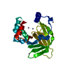
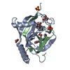
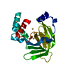
- PDB-1txl: Crystal structure of metal-binding protein yodA from E. coli, Pfa... -
+
Open data
ID or keywords:
Loading...
-
Basic information
Entry
Database: PDB / ID: 1txl
Title
Crystal structure of metal-binding protein yodA from E. coli, Pfam DUF149
Components
Metal-binding protein yodA
Keywords
structural genomics / unknown function / yodA / E.coli / new fold / PSI / Protein Structure Initiative / New York SGX Research Center for Structural Genomics / NYSGXRC
Function / homology
Function and homology information
cellular response to zinc ion starvation / intracellular zinc ion homeostasis / cadmium ion binding / cellular response to cadmium ion / cellular response to hydrogen peroxide / outer membrane-bounded periplasmic space / zinc ion binding / metal ion binding / cytosol Similarity search - Function
Method to determine structure: MAD / Resolution: 1.7→50 Å / Cross valid method: THROUGHOUT / σ(F): 0 / Stereochemistry target values: Engh & Huber Details: A Zn atom coordinating to HIS166, HIS175 and HIS177 has been located in the putative active-site cavity. Followed by Zn, continuous electron density has been observed. This density could ...Details: A Zn atom coordinating to HIS166, HIS175 and HIS177 has been located in the putative active-site cavity. Followed by Zn, continuous electron density has been observed. This density could accomodate a branched carbohydrate chain and a PO4 head group. A probable substrate suspected to be present in the structure has not been added to the model because it is not known for sure. Water molecules located around the Zn atom in the active site cavity have to be treated with care. Water molecule 218 may be replaced with a PO4.
Rfactor
Num. reflection
Selection details
Rfree
0.2522
973
RANDOM
Rwork
0.2282
-
-
obs
0.2282
20185
-
all
-
20185
-
Refinement step
Cycle: LAST / Resolution: 1.7→50 Å
Protein
Nucleic acid
Ligand
Solvent
Total
Num. atoms
1537
0
1
224
1762
Refine LS restraints
Refine-ID
Type
Dev ideal
X-RAY DIFFRACTION
c_angle_deg
1.2451
X-RAY DIFFRACTION
c_bond_d
0.0065
+
About Yorodumi
-
News
-
Feb 9, 2022. New format data for meta-information of EMDB entries
New format data for meta-information of EMDB entries
Version 3 of the EMDB header file is now the official format.
The previous official version 1.9 will be removed from the archive.
In the structure databanks used in Yorodumi, some data are registered as the other names, "COVID-19 virus" and "2019-nCoV". Here are the details of the virus and the list of structure data.
Jan 31, 2019. EMDB accession codes are about to change! (news from PDBe EMDB page)
EMDB accession codes are about to change! (news from PDBe EMDB page)
The allocation of 4 digits for EMDB accession codes will soon come to an end. Whilst these codes will remain in use, new EMDB accession codes will include an additional digit and will expand incrementally as the available range of codes is exhausted. The current 4-digit format prefixed with “EMD-” (i.e. EMD-XXXX) will advance to a 5-digit format (i.e. EMD-XXXXX), and so on. It is currently estimated that the 4-digit codes will be depleted around Spring 2019, at which point the 5-digit format will come into force.
The EM Navigator/Yorodumi systems omit the EMD- prefix.
Related info.:Q: What is EMD? / ID/Accession-code notation in Yorodumi/EM Navigator
Yorodumi is a browser for structure data from EMDB, PDB, SASBDB, etc.
This page is also the successor to EM Navigator detail page, and also detail information page/front-end page for Omokage search.
The word "yorodu" (or yorozu) is an old Japanese word meaning "ten thousand". "mi" (miru) is to see.
Related info.:EMDB / PDB / SASBDB / Comparison of 3 databanks / Yorodumi Search / Aug 31, 2016. New EM Navigator & Yorodumi / Yorodumi Papers / Jmol/JSmol / Function and homology information / Changes in new EM Navigator and Yorodumi
 Movie
Movie Controller

 Yorodumi
 Open data
 Basic information
 Components
 Keywords
 Function and homology information
 X-RAY DIFFRACTION /
  Authors
 Citation
 Structure visualization
 Downloads & links
 Other downloads
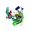


 PDBj
PDBj

 Assembly
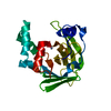

 Mass: 65.409 Da / Num. of mol.: 1 / Source method: obtained synthetically / Formula: Zn
 Mass: 18.015 Da / Num. of mol.: 224 / Source method: isolated from a natural source / Formula: H2O
 Sample preparation
 Processing