 Movie
Movie Controller

[English] 日本語
 Yorodumi
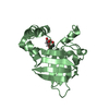
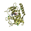
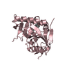
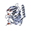
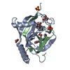
- PDB-5xm5: Crystal structure of Zinc binding protein ZinT at 1.49 Angstrom f... -
+ Open data

- Basic information
| Entry | Database: PDB / ID: 5xm5 | ||||||||||||
|---|---|---|---|---|---|---|---|---|---|---|---|---|---|
| Title | Crystal structure of Zinc binding protein ZinT at 1.49 Angstrom from E. coli | ||||||||||||
 Components Components | Metal-binding protein ZinT | ||||||||||||
 Keywords Keywords | METAL BINDING PROTEIN / Zinc binding protein | ||||||||||||
| Function / homology |  Function and homology information Function and homology informationcellular response to zinc ion starvation / intracellular zinc ion homeostasis / cadmium ion binding / cellular response to cadmium ion / cellular response to hydrogen peroxide / outer membrane-bounded periplasmic space / zinc ion binding / metal ion binding / cytosol Similarity search - Function | ||||||||||||
| Biological species |  | ||||||||||||
| Method |  X-RAY DIFFRACTION / SYNCHROTRON / MOLECULAR REPLACEMENT / Resolution: 1.493 Å X-RAY DIFFRACTION / SYNCHROTRON / MOLECULAR REPLACEMENT / Resolution: 1.493 Å | ||||||||||||
 Authors Authors | Chen, J. / Wang, L. / Guo, J. / Xu, Y. | ||||||||||||
| Funding support |  China, 3items China, 3items
| ||||||||||||
 Citation Citation | Journal: To Be Published Title: Crystal structure of Zinc binding protein ZinT at 1.49 Angstrom from E. coli Authors: Chen, J. / Wang, L. / Guo, J. / Xu, Y. | ||||||||||||
| History |
|
- Structure visualization
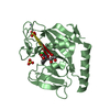
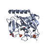
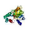
| Structure viewer | Molecule: MolmilJmol/JSmol |
|---|
- Downloads & links
-Download
| PDBx/mmCIF format | 5xm5.cif.gz | 233 KB | Display | PDBx/mmCIF format |
|---|---|---|---|---|
| PDB format | pdb5xm5.ent.gz | 191 KB | Display | PDB format |
| PDBx/mmJSON format | 5xm5.json.gz | Tree view | PDBx/mmJSON format | |
| Others |  Other downloads Other downloads |
-Validation report
| Arichive directory | https://data.pdbj.org/pub/pdb/validation_reports/xm/5xm5ftp://data.pdbj.org/pub/pdb/validation_reports/xm/5xm5 | HTTPS FTP |
|---|
-Related structure data
| Related structure data |  4aw8S S: Starting model for refinement |
|---|---|
| Similar structure data |


-Links
 PDBj
PDBj

- Assembly
| Deposited unit | 
| ||||||||
|---|---|---|---|---|---|---|---|---|---|
| 1 | 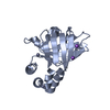
| ||||||||
| 2 | 
| ||||||||
| Unit cell |
|
-Components
| #1: Protein | Mass: 22373.879 Da / Num. of mol.: 2 / Fragment: UNP residue 24-216 Source method: isolated from a genetically manipulated source Source: (gene. exp.) Strain: K12 / Gene: zinT, yodA, b1973, JW1956 Production host: References: UniProt: P76344 #2: Chemical | ChemComp-ZN /   Mass: 65.409 Da / Num. of mol.: 6 / Source method: obtained synthetically / Formula: Zn Mass: 65.409 Da / Num. of mol.: 6 / Source method: obtained synthetically / Formula: Zn#3: Chemical | ChemComp-NA /   Mass: 22.990 Da / Num. of mol.: 5 / Source method: obtained synthetically / Formula: Na Mass: 22.990 Da / Num. of mol.: 5 / Source method: obtained synthetically / Formula: Na#4: Water | ChemComp-HOH / |  Mass: 18.015 Da / Num. of mol.: 519 / Source method: isolated from a natural source / Formula: H2O Mass: 18.015 Da / Num. of mol.: 519 / Source method: isolated from a natural source / Formula: H2OHas protein modification | Y | |
|---|
-Experimental details
-Experiment
| Experiment | Method: X-RAY DIFFRACTION / Number of used crystals: 1 |
|---|
- Sample preparation
| Crystal | Density Matthews: 2.21 Å3/Da / Density % sol: 44.46 % |
|---|---|
| Crystal grow | Temperature: 288 K / Method: vapor diffusion, sitting drop / pH: 4.6 Details: 22.5%(w/v) polyethylene glycol 4000, 0.135M ammonium acetate, and 0.1M sodium acetate trihydrate (pH 4.6) |
-Data collection
| Diffraction | Mean temperature: 100 K / Ambient temp details: Liquid nitrogen |
|---|---|
| Diffraction source | Source: SYNCHROTRON / Type: RIGAKU / Wavelength: 0.979 Å |
| Detector | Type: ADSC QUANTUM 315r / Detector: CCD / Date: Nov 3, 2016 |
| Radiation | Protocol: SINGLE WAVELENGTH / Monochromatic (M) / Laue (L): M / Scattering type: x-ray |
| Radiation wavelength | Wavelength: 0.979 Å / Relative weight: 1 |
| Reflection | Resolution: 1.493→31.228 Å / Num. obs: 56913 / % possible obs: 90.5 % / Redundancy: 2.2 % / Net I/σ(I): 13.319 |
| Reflection shell | Resolution: 1.49→1.52 Å / Redundancy: 9.5 % / Mean I/σ(I) obs: 16.37 / Num. unique obs: 54181 / % possible all: 90.5 |
- Processing
| Software |
| |||||||||||||||||||||||||||||||||||||||||||||||||||||||||||||||||||||||||||||||||||||||||||||||||||||||||
|---|---|---|---|---|---|---|---|---|---|---|---|---|---|---|---|---|---|---|---|---|---|---|---|---|---|---|---|---|---|---|---|---|---|---|---|---|---|---|---|---|---|---|---|---|---|---|---|---|---|---|---|---|---|---|---|---|---|---|---|---|---|---|---|---|---|---|---|---|---|---|---|---|---|---|---|---|---|---|---|---|---|---|---|---|---|---|---|---|---|---|---|---|---|---|---|---|---|---|---|---|---|---|---|---|---|---|
| Refinement | Method to determine structure: MOLECULAR REPLACEMENT Starting model: 4aw8 Resolution: 1.493→31.228 Å / SU ML: 0.21 / Cross valid method: FREE R-VALUE / σ(F): 1.33 / Phase error: 31.18
| |||||||||||||||||||||||||||||||||||||||||||||||||||||||||||||||||||||||||||||||||||||||||||||||||||||||||
| Solvent computation | Shrinkage radii: 0.9 Å / VDW probe radii: 1.11 Å | |||||||||||||||||||||||||||||||||||||||||||||||||||||||||||||||||||||||||||||||||||||||||||||||||||||||||
| Refinement step | Cycle: LAST / Resolution: 1.493→31.228 Å
| |||||||||||||||||||||||||||||||||||||||||||||||||||||||||||||||||||||||||||||||||||||||||||||||||||||||||
| Refine LS restraints |
| |||||||||||||||||||||||||||||||||||||||||||||||||||||||||||||||||||||||||||||||||||||||||||||||||||||||||
| LS refinement shell |
|
