 Movie
Movie Controller


+ Open data

- Basic information
| Entry | Database: PDB / ID: 6lm2 | ||||||
|---|---|---|---|---|---|---|---|
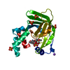
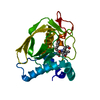
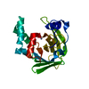
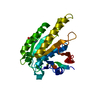
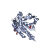
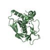
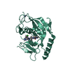
| Title | Crystal structure of Zinc binding protein ZinT from E. coli | ||||||
 Components Components | Metal-binding protein ZinT | ||||||
 Keywords Keywords | METAL BINDING PROTEIN / Zinc binding protein ZinT from E. coli | ||||||
| Function / homology |  Function and homology information Function and homology informationcellular response to zinc ion starvation / intracellular zinc ion homeostasis / cadmium ion binding / cellular response to cadmium ion / cellular response to hydrogen peroxide / outer membrane-bounded periplasmic space / zinc ion binding / metal ion binding / cytosol Similarity search - Function | ||||||
| Biological species | Escherichia coli 'BL21-GoldpLysS AG' | ||||||
| Method |  X-RAY DIFFRACTION / SYNCHROTRON / MOLECULAR REPLACEMENT / Resolution: 2.13008410001 Å X-RAY DIFFRACTION / SYNCHROTRON / MOLECULAR REPLACEMENT / Resolution: 2.13008410001 Å | ||||||
 Authors Authors | Xiang, L. / Zhang, G. / Zhou, J. | ||||||
 Citation Citation | Journal: To Be Published Title: Crystal structure of Zinc binding protein ZinT from E. coli Authors: Xiang, L. / Zhang, G. / Zhou, J. | ||||||
| History |
|
- Structure visualization
| Structure viewer | Molecule: MolmilJmol/JSmol |
|---|
- Downloads & links
-Download
| PDBx/mmCIF format | 6lm2.cif.gz | 108.6 KB | Display | PDBx/mmCIF format |
|---|---|---|---|---|
| PDB format | pdb6lm2.ent.gz | 69.7 KB | Display | PDB format |
| PDBx/mmJSON format | 6lm2.json.gz | Tree view | PDBx/mmJSON format | |
| Others |  Other downloads Other downloads |
-Validation report
| Arichive directory | https://data.pdbj.org/pub/pdb/validation_reports/lm/6lm2ftp://data.pdbj.org/pub/pdb/validation_reports/lm/6lm2 | HTTPS FTP |
|---|
-Related structure data
| Related structure data | 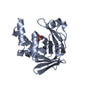 5yxcS S: Starting model for refinement |
|---|---|
| Similar structure data |


-Links
 PDBj
PDBj

- Assembly
| Deposited unit | 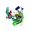
| ||||||||||||
|---|---|---|---|---|---|---|---|---|---|---|---|---|---|
| 1 |
| ||||||||||||
| Unit cell |
| ||||||||||||
| Components on special symmetry positions |
|
-Components
| #1: Protein | Mass: 21292.750 Da / Num. of mol.: 1 / Source method: isolated from a natural source Source: (natural)  Strain: 'BL21-Gold(DE3)pLysS AG' / References: UniProt: A0A3Z9V4E1, UniProt: P76344*PLUS | ||||||
|---|---|---|---|---|---|---|---|
| #2: Chemical | ChemComp-ZN /   Mass: 65.409 Da / Num. of mol.: 8 / Source method: obtained synthetically / Formula: Zn / Feature type: SUBJECT OF INVESTIGATION Mass: 65.409 Da / Num. of mol.: 8 / Source method: obtained synthetically / Formula: Zn / Feature type: SUBJECT OF INVESTIGATION#3: Water | ChemComp-HOH / |  Mass: 18.015 Da / Num. of mol.: 38 / Source method: isolated from a natural source / Formula: H2O Mass: 18.015 Da / Num. of mol.: 38 / Source method: isolated from a natural source / Formula: H2OHas ligand of interest | Y | Has protein modification | Y | |
-Experimental details
-Experiment
| Experiment | Method: X-RAY DIFFRACTION / Number of used crystals: 1 |
|---|
- Sample preparation
| Crystal | Density Matthews: 2.81 Å3/Da / Density % sol: 56.15 % |
|---|---|
| Crystal grow | Temperature: 289 K / Method: vapor diffusion, sitting drop Details: 0.2 M Zinc acetate, 0.1 M Sodium acetate pH 4.5, 10 %(w/v) PEG 3000 |
-Data collection
| Diffraction | Mean temperature: 100 K / Serial crystal experiment: N |
|---|---|
| Diffraction source | Source: SYNCHROTRON / Site: SSRF  / Beamline: BL18U1 / Wavelength: 0.9793 Å / Beamline: BL18U1 / Wavelength: 0.9793 Å |
| Detector | Type: DECTRIS PILATUS3 S 6M / Detector: PIXEL / Date: Nov 25, 2019 |
| Radiation | Protocol: SINGLE WAVELENGTH / Monochromatic (M) / Laue (L): M / Scattering type: x-ray |
| Radiation wavelength | Wavelength: 0.9793 Å / Relative weight: 1 |
| Reflection | Resolution: 2.13→45.13 Å / Num. obs: 14365 / % possible obs: 99.9 % / Redundancy: 24.6 % / Biso Wilson estimate: 36.7608797957 Å2 / CC1/2: 0.998 / Net I/σ(I): 17.4 |
| Reflection shell | Resolution: 2.13→2.19 Å / Num. unique obs: 1138 / CC1/2: 0.974 |
- Processing
| Software |
| ||||||||||||||||||||||||||||||||||||||||||
|---|---|---|---|---|---|---|---|---|---|---|---|---|---|---|---|---|---|---|---|---|---|---|---|---|---|---|---|---|---|---|---|---|---|---|---|---|---|---|---|---|---|---|---|
| Refinement | Method to determine structure: MOLECULAR REPLACEMENT Starting model: 5YXC Resolution: 2.13008410001→38.355 Å / SU ML: 0.257544649448 / Cross valid method: FREE R-VALUE / σ(F): 1.34349736043 / Phase error: 31.5680159947
| ||||||||||||||||||||||||||||||||||||||||||
| Solvent computation | Shrinkage radii: 0.9 Å / VDW probe radii: 1.11 Å | ||||||||||||||||||||||||||||||||||||||||||
| Displacement parameters | Biso mean: 50.7763735099 Å2 | ||||||||||||||||||||||||||||||||||||||||||
| Refinement step | Cycle: LAST / Resolution: 2.13008410001→38.355 Å
| ||||||||||||||||||||||||||||||||||||||||||
| Refine LS restraints |
| ||||||||||||||||||||||||||||||||||||||||||
| LS refinement shell |
| ||||||||||||||||||||||||||||||||||||||||||
| Refinement TLS params. | Method: refined / Origin x: 5.83049992863 Å / Origin y: 26.2568392815 Å / Origin z: 67.2037012503 Å
| ||||||||||||||||||||||||||||||||||||||||||
| Refinement TLS group | Selection details: all |
