 Movie
Movie Controller


+ Open data

- Basic information
| Entry | Database: PDB / ID: 1qmv | ||||||
|---|---|---|---|---|---|---|---|
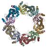
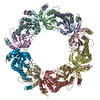
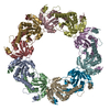
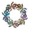
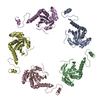
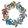
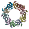
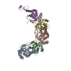
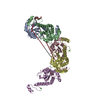
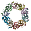
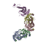
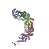
| Title | thioredoxin peroxidase B from red blood cells | ||||||
 Components Components | PEROXIREDOXIN-2 | ||||||
 Keywords Keywords | OXIDOREDUCTASE / SULPHINIC ACID / THIOREDOXIN | ||||||
| Function / homology |  Function and homology information Function and homology informationrespiratory burst involved in inflammatory response / negative regulation of T cell differentiation / leukocyte activation / regulation of hydrogen peroxide metabolic process / thioredoxin-dependent peroxiredoxin / negative regulation of lipopolysaccharide-mediated signaling pathway / thioredoxin peroxidase activity / defense response to tumor cell / Detoxification of Reactive Oxygen Species / Deregulated CDK5 triggers multiple neurodegenerative pathways in Alzheimer's disease models ...respiratory burst involved in inflammatory response / negative regulation of T cell differentiation / leukocyte activation / regulation of hydrogen peroxide metabolic process / thioredoxin-dependent peroxiredoxin / negative regulation of lipopolysaccharide-mediated signaling pathway / thioredoxin peroxidase activity / defense response to tumor cell / Detoxification of Reactive Oxygen Species / Deregulated CDK5 triggers multiple neurodegenerative pathways in Alzheimer's disease models / antioxidant activity / T cell homeostasis / positive regulation of blood coagulation / T cell proliferation / negative regulation of extrinsic apoptotic signaling pathway in absence of ligand / extrinsic apoptotic signaling pathway / removal of superoxide radicals / cell redox homeostasis / thymus development / hydrogen peroxide catabolic process / TP53 Regulates Metabolic Genes / cellular response to oxidative stress / response to oxidative stress / regulation of apoptotic process / response to lipopolysaccharide / positive regulation of MAPK cascade / negative regulation of apoptotic process / extracellular exosome / cytoplasm / cytosol Similarity search - Function | ||||||
| Biological species |  HOMO SAPIENS (human) HOMO SAPIENS (human) | ||||||
| Method |  X-RAY DIFFRACTION / SYNCHROTRON / MOLECULAR REPLACEMENT / Resolution: 1.7 Å X-RAY DIFFRACTION / SYNCHROTRON / MOLECULAR REPLACEMENT / Resolution: 1.7 Å | ||||||
 Authors Authors | Isupov, M.N. / Littlechild, J.A. / Lebedev, A.A. / Errington, N. / Vagin, A.A. / Schroder, E. | ||||||
 Citation Citation | Journal: Structure / Year: 2000 Title: Crystal Structure of Decameric 2-Cys Peroxiredoxin from Human Erythrocytes at 1.7 A Resolution. Authors: Schroder, E. / Littlechild, J.A. / Lebedev, A.A. / Errington, N. / Vagin, A.A. / Isupov, M.N. #1: Journal: Acta Crystallogr.,Sect.D / Year: 1999 Title: Crystallisation and Preliminary X-Ray Analysis of Human Thioredoxin Peroxidase B from Red Blood Cells Authors: Schroder, E. / Isupov, M.N. / Naran, A. / Littlechild, J.A. #2: Journal: Protein Sci. / Year: 1998 Title: Evidence that Peroxiredoxins are Novel Members of the Thioredoxin Fold Superfamily Authors: Schroder, E. / Ponting, C.P. #3: Journal: Biochim.Biophys.Acta / Year: 1998 Title: Porcine Natural Killer Enhancing Factor-B: Oligomerization and Identification as a Calpain Substrate in-Vitro Authors: Schroder, E. / Willis, A.C. / Ponting, C.P. | ||||||
| History |
|
- Structure visualization
| Structure viewer | Molecule: MolmilJmol/JSmol |
|---|
- Downloads & links
-Download
| PDBx/mmCIF format | 1qmv.cif.gz | 425.7 KB | Display | PDBx/mmCIF format |
|---|---|---|---|---|
| PDB format | pdb1qmv.ent.gz | 352.2 KB | Display | PDB format |
| PDBx/mmJSON format | 1qmv.json.gz | Tree view | PDBx/mmJSON format | |
| Others |  Other downloads Other downloads |
-Validation report
| Arichive directory | https://data.pdbj.org/pub/pdb/validation_reports/qm/1qmvftp://data.pdbj.org/pub/pdb/validation_reports/qm/1qmv | HTTPS FTP |
|---|
-Related structure data
| Related structure data |  1prxS S: Starting model for refinement |
|---|---|
| Similar structure data |

-Links
 PDBj
PDBj


- Assembly
| Deposited unit | 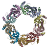
| ||||||||||||||||||||||||||||||||||||||||
|---|---|---|---|---|---|---|---|---|---|---|---|---|---|---|---|---|---|---|---|---|---|---|---|---|---|---|---|---|---|---|---|---|---|---|---|---|---|---|---|---|---|
| 1 |
| ||||||||||||||||||||||||||||||||||||||||
| Unit cell |
| ||||||||||||||||||||||||||||||||||||||||
| Noncrystallographic symmetry (NCS) | NCS oper:
|
-Components
| #1: Protein | Mass: 21862.715 Da / Num. of mol.: 10 / Source method: isolated from a natural source / Details: PURIFIED FROM ONE MONTH OLD BLOOD / Source: (natural) HOMO SAPIENS (human) / Cell: ERYTHROCYTE / Cellular location: CYTOPLASM / Organ: BLOOD / References: UniProt: P32119, peroxiredoxin#2: Water | ChemComp-HOH / |  Mass: 18.015 Da / Num. of mol.: 2194 / Source method: isolated from a natural source / Formula: H2O Mass: 18.015 Da / Num. of mol.: 2194 / Source method: isolated from a natural source / Formula: H2OHas protein modification | Y | |
|---|
-Experimental details
-Experiment
| Experiment | Method: X-RAY DIFFRACTION / Number of used crystals: 1 |
|---|
- Sample preparation
| Crystal | Density Matthews: 2.4 Å3/Da / Density % sol: 49 % | |||||||||||||||||||||||||||||||||||||||||||||
|---|---|---|---|---|---|---|---|---|---|---|---|---|---|---|---|---|---|---|---|---|---|---|---|---|---|---|---|---|---|---|---|---|---|---|---|---|---|---|---|---|---|---|---|---|---|---|
| Crystal grow | pH: 7.5 Details: 16 % (V/V) PEG 400, 100 MM TRIS-HCL, PH 7.5, 10 % (V/V) 2-PROPANOL, 10 MM DITHIOERYTHREITOL | |||||||||||||||||||||||||||||||||||||||||||||
| Crystal grow | *PLUS Temperature: 290 K / Method: vapor diffusion, hanging dropDetails: drop consists of equal volume of protein and reservoir solutions | |||||||||||||||||||||||||||||||||||||||||||||
| Components of the solutions | *PLUS
|
-Data collection
| Diffraction | Mean temperature: 100 K |
|---|---|
| Diffraction source | Source: SYNCHROTRON / Site: EMBL/DESY, HAMBURG  / Beamline: X11 / Wavelength: 0.9076 / Beamline: X11 / Wavelength: 0.9076 |
| Detector | Type: MARRESEARCH / Detector: IMAGE PLATE / Date: Jul 15, 1998 |
| Radiation | Protocol: SINGLE WAVELENGTH / Monochromatic (M) / Laue (L): M / Scattering type: x-ray |
| Radiation wavelength | Wavelength: 0.9076 Å / Relative weight: 1 |
| Reflection | Resolution: 1.7→20 Å / Num. obs: 228010 / % possible obs: 99.6 % / Redundancy: 4.2 % / Biso Wilson estimate: 34.1 Å2 / Rsym value: 0.057 / Net I/σ(I): 18.8 |
| Reflection shell | Resolution: 1.7→1.73 Å / Redundancy: 3 % / Mean I/σ(I) obs: 1.7 / Rsym value: 0.493 / % possible all: 93.6 |
| Reflection | *PLUS Rmerge(I) obs: 0.057 |
| Reflection shell | *PLUS % possible obs: 93.6 % / Redundancy: 2.3 % / Rmerge(I) obs: 0.481 / Mean I/σ(I) obs: 2.07 |
- Processing
| Software |
| ||||||||||||||||||||||||||||||||||||||||||||||||||||||||||||||||||||||||||||||||||||
|---|---|---|---|---|---|---|---|---|---|---|---|---|---|---|---|---|---|---|---|---|---|---|---|---|---|---|---|---|---|---|---|---|---|---|---|---|---|---|---|---|---|---|---|---|---|---|---|---|---|---|---|---|---|---|---|---|---|---|---|---|---|---|---|---|---|---|---|---|---|---|---|---|---|---|---|---|---|---|---|---|---|---|---|---|---|
| Refinement | Method to determine structure: MOLECULAR REPLACEMENT Starting model: PDB ENTRY 1PRX Resolution: 1.7→20 Å / SU B: 2.67 / SU ML: 0.084 / Cross valid method: THROUGHOUT / σ(F): 0 / ESU R: 0.1106 / ESU R Free: 0.122
| ||||||||||||||||||||||||||||||||||||||||||||||||||||||||||||||||||||||||||||||||||||
| Displacement parameters | Biso mean: 32.7 Å2
| ||||||||||||||||||||||||||||||||||||||||||||||||||||||||||||||||||||||||||||||||||||
| Refinement step | Cycle: LAST / Resolution: 1.7→20 Å
| ||||||||||||||||||||||||||||||||||||||||||||||||||||||||||||||||||||||||||||||||||||
| Refine LS restraints |
|
