 Movie
Movie Controller


+ Open data

- Basic information
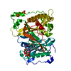
| Entry | Database: PDB / ID: 1prx | ||||||
|---|---|---|---|---|---|---|---|
| Title | HORF6 A NOVEL HUMAN PEROXIDASE ENZYME | ||||||
 Components Components | HORF6 | ||||||
 Keywords Keywords | ANTIOXIDANT / PEROXIREDOXIN / HORF6 / HYDROGEN PEROXIDE / REDOX REGULATION / CELLULAR SIGNALING | ||||||
| Function / homology |  Function and homology information Function and homology information1-acylglycerophosphocholine O-acyltransferase / 1-acylglycerophosphocholine O-acyltransferase activity / glycerophospholipid catabolic process / glutathione-dependent peroxiredoxin / : / A2-type glycerophospholipase activity / peroxiredoxin activity / glutathione peroxidase activity / positive regulation of mRNA splicing, via spliceosome / phospholipase A2 ...1-acylglycerophosphocholine O-acyltransferase / 1-acylglycerophosphocholine O-acyltransferase activity / glycerophospholipid catabolic process / glutathione-dependent peroxiredoxin / : / A2-type glycerophospholipase activity / peroxiredoxin activity / glutathione peroxidase activity / positive regulation of mRNA splicing, via spliceosome / phospholipase A2 / cellular oxidant detoxification / Detoxification of Reactive Oxygen Species / cell redox homeostasis / hydrogen peroxide catabolic process / peroxidase activity / azurophil granule lumen / response to oxidative stress / cadherin binding / Neutrophil degranulation / ubiquitin protein ligase binding / perinuclear region of cytoplasm / : / extracellular exosome / extracellular region / membrane / identical protein binding / nucleus / cytoplasm / cytosol Similarity search - Function | ||||||
| Biological species |  Homo sapiens (human) Homo sapiens (human) | ||||||
| Method |  X-RAY DIFFRACTION / MIR / Resolution: 2 Å X-RAY DIFFRACTION / MIR / Resolution: 2 Å | ||||||
 Authors Authors | Choi, H.-J. / Kang, S.W. / Yang, C.-H. / Rhee, S.G. / Ryu, S.-E. | ||||||
 Citation Citation | Journal: Nat.Struct.Biol. / Year: 1998 Title: Crystal structure of a novel human peroxidase enzyme at 2.0 A resolution. Authors: Choi, H.J. / Kang, S.W. / Yang, C.H. / Rhee, S.G. / Ryu, S.E. | ||||||
| History |
|
- Structure visualization
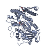
| Structure viewer | Molecule: MolmilJmol/JSmol |
|---|
- Downloads & links
-Download
| PDBx/mmCIF format | 1prx.cif.gz | 97.4 KB | Display | PDBx/mmCIF format |
|---|---|---|---|---|
| PDB format | pdb1prx.ent.gz | 74.6 KB | Display | PDB format |
| PDBx/mmJSON format | 1prx.json.gz | Tree view | PDBx/mmJSON format | |
| Others |  Other downloads Other downloads |
-Validation report
| Arichive directory | https://data.pdbj.org/pub/pdb/validation_reports/pr/1prxftp://data.pdbj.org/pub/pdb/validation_reports/pr/1prx | HTTPS FTP |
|---|
-Related structure data
| Similar structure data |
|---|


-Links
 PDBj
PDBj


- Assembly
| Deposited unit | 
| ||||||||
|---|---|---|---|---|---|---|---|---|---|
| 1 |
| ||||||||
| Unit cell |
| ||||||||
| Noncrystallographic symmetry (NCS) | NCS oper: (Code: given Matrix: (-0.99988, -0.01523, 0.00341), Vector: |
-Components
| #1: Protein | Mass: 25067.904 Da / Num. of mol.: 2 / Mutation: C91S Source method: isolated from a genetically manipulated source Source: (gene. exp.) Homo sapiens (human) / Production host:  #2: Water | ChemComp-HOH / |  Mass: 18.015 Da / Num. of mol.: 112 / Source method: isolated from a natural source / Formula: H2O Mass: 18.015 Da / Num. of mol.: 112 / Source method: isolated from a natural source / Formula: H2OHas protein modification | Y | |
|---|
-Experimental details
-Experiment
| Experiment | Method: X-RAY DIFFRACTION / Number of used crystals: 8 |
|---|
- Sample preparation
| Crystal | Density Matthews: 2.13 Å3/Da / Density % sol: 42.23 % | ||||||||||||||||||||
|---|---|---|---|---|---|---|---|---|---|---|---|---|---|---|---|---|---|---|---|---|---|
| Crystal grow | pH: 6.5 / Details: pH 6.5 | ||||||||||||||||||||
| Crystal | *PLUS Density % sol: 43 % | ||||||||||||||||||||
| Crystal grow | *PLUS Temperature: 295 K / Method: vapor diffusion, hanging drop | ||||||||||||||||||||
| Components of the solutions | *PLUS
|
-Data collection
| Diffraction | Mean temperature: 295 K |
|---|---|
| Diffraction source | Source: ROTATING ANODE / Type: RIGAKU RUH3R / Wavelength: 1.5418 |
| Detector | Type: RIGAKU / Detector: IMAGE PLATE / Date: Aug 1, 1996 |
| Radiation | Monochromator: GRAPHITE(002) / Monochromatic (M) / Laue (L): M / Scattering type: x-ray |
| Radiation wavelength | Wavelength: 1.5418 Å / Relative weight: 1 |
| Reflection | Resolution: 2→8 Å / Num. obs: 24439 / % possible obs: 93.7 % / Observed criterion σ(I): 1 / Redundancy: 3 % / Rmerge(I) obs: 0.07 / Rsym value: 0.07 / Net I/σ(I): 17 |
| Reflection shell | Resolution: 2→2.25 Å / Redundancy: 3 % / Rmerge(I) obs: 0.189 / Mean I/σ(I) obs: 6.1 / Rsym value: 0.189 / % possible all: 91.4 |
| Reflection | *PLUS Highest resolution: 2 Å / Lowest resolution: 8 Å |
| Reflection shell | *PLUS % possible obs: 91.4 % |
- Processing
| Software |
| ||||||||||||||||||||||||||||||||||||||||||||||||||||||||||||
|---|---|---|---|---|---|---|---|---|---|---|---|---|---|---|---|---|---|---|---|---|---|---|---|---|---|---|---|---|---|---|---|---|---|---|---|---|---|---|---|---|---|---|---|---|---|---|---|---|---|---|---|---|---|---|---|---|---|---|---|---|---|
| Refinement | Method to determine structure: MIR / Resolution: 2→8 Å / Data cutoff high absF: 10000000 / Data cutoff low absF: 0 / Isotropic thermal model: RESTRAINED / Cross valid method: THROUGHOUT / σ(F): 2
| ||||||||||||||||||||||||||||||||||||||||||||||||||||||||||||
| Displacement parameters | Biso mean: 20.91 Å2 | ||||||||||||||||||||||||||||||||||||||||||||||||||||||||||||
| Refinement step | Cycle: LAST / Resolution: 2→8 Å
| ||||||||||||||||||||||||||||||||||||||||||||||||||||||||||||
| Refine LS restraints |
| ||||||||||||||||||||||||||||||||||||||||||||||||||||||||||||
| LS refinement shell | Resolution: 2→2.25 Å | ||||||||||||||||||||||||||||||||||||||||||||||||||||||||||||
| Xplor file |
| ||||||||||||||||||||||||||||||||||||||||||||||||||||||||||||
| Software | *PLUS Name: X-PLOR / Version: 3.8 / Classification: refinement | ||||||||||||||||||||||||||||||||||||||||||||||||||||||||||||
| Refinement | *PLUS | ||||||||||||||||||||||||||||||||||||||||||||||||||||||||||||
| Solvent computation | *PLUS | ||||||||||||||||||||||||||||||||||||||||||||||||||||||||||||
| Displacement parameters | *PLUS | ||||||||||||||||||||||||||||||||||||||||||||||||||||||||||||
| Refine LS restraints | *PLUS
|
